
System and method for implantation of lead and electrodes to the endopelvic portion of the pelvic nerves and connection cable for electrode with direction marker

a technology of pelvic nerve and pelvis, which is applied in the direction of internal electrodes, artificial respiration, therapy, etc., can solve the problems of increased bladder pressure, decreased urethra function, and adverse effects of pelvic floor disorders on the health and quality of life of millions of people, so as to prevent decubitus ulcers, minimize decubitus ulcers, and prevent decubitus ulcers

Inactive Publication Date: 2017-02-16
POSSOVER MARC
View PDF6 Cites 3 Cited by
  • Summary
  • Abstract
  • Description
  • Claims
  • Application Information

AI Technical Summary

Benefits of technology

The patent text discusses the benefits of PN stimulation and its use in treating erectile dysfunction and osteoporosis. The technical effects include improved erectile function and increased blood supply to bones, which prevents osteoporosis and treats it effectively.

Problems solved by technology

Pelvic floor disorders adversely affect the health and quality of life of millions of people.
These activities and events cause an increase in bladder pressure resulting in loss of urine due to inadequate contraction of the sphincter muscle around the outlet of the bladder.
Many IC patients also experience headaches as well as gastrointestinal and skin problems.
Fecal incontinence is the inability to control your bowel movements, causing stool (feces) to leak unexpectedly from your rectum.
Common causes of fecal incontinence include constipation, diarrhea, and muscle or nerve damage.
An erection cannot be sustained without sufficient blood flow into and entrapment within the erectile bodies of the penis, and vascular related erectile dysfunctions can be due to a malfunction of either the arterial or the venous system.
Pharmacotherapy appears to moderate the incidence of UI episodes, but not eliminate them.
Limitations of medical treatment may be limited efficacy over time, but first of all side effects such as dry mouth, dry eyes, dry vagina, blurred vision, cardiac side effects, such as palpitations and arrhythmia, drowsiness, urinary retention, weight gain, hypertension and constipation, which have proven difficult for some individuals to tolerate.
This surgical procedure near the spine is complex and requires the skills of specialized medical personnel.
In terms of outcomes, the modality has demonstrated limited effectiveness.
This modality has also demonstrated limited reliability.
In combinations of urinary and faecal disorders, because sacral nerve stimulation does not permit stimulation and / or neuromodulation of all pudendal fibers, it is difficult to treat urinary and faecal disorders with the same effectiveness.
The clinical effectiveness of this modality has not been proved; the main problem is high rate of migration of the implant away from the pudendal nerves, with risk of migration being increased by sitting position, gluteal muscle activation and in women, sexual activities.
Furthermore, this technique is too restrictive since it enables only treatment of urinary dysfunctions but not faecal dysfunctions or all pelvic pain situations (vulvodynia, pudendal neuralgia, etc.).
A technology of this type, assumed to be category-defining, is complicated however in terms of handling and implementation: not only are considerable demands placed on the surgical knowledge or surgical capability of the operator in question, but the substantially parallel alignment, required by the known technology, between optical observation axis on the one hand (the visual control or controllability by means of the endoscope) and the feed of the electrode wire through the endoscopic working shaft on the other hand is also unfavorable for exact alignment and positioning specifically of the critical nerve contact portion at the end of the electrode wire.
In other words, simple and reliably positioned handling of the electrode wire at the site of implantation in the human inner pelvic region under optical control of the endoscope is impeded especially with orthogonally running geometries, which further increases the demands placed on the operator.
A further problem with this device known from the prior art lies in the fact that, with a wire electrode implanted via the working channel of an endoscope, said wire electrode (once the endoscope has been removed, whereby the electrode is then left at the site of implantation) protrudes via its connection portion opposite the nerve contact portion from the bodily access point used for the endoscope (typically arranged in the abdominal region, for example the navel).
In order to then connect this connection portion of the electrode wire to a signal generation source (which typically is also implanted beneath the patient's skin), it is necessary to lay or surgically pass the connection-side end of the electrode wire in the superficial bodily region, which additionally increases the complexity of the procedure and subjects the patient to further potential stress.
It can happen that the operator pulls the connection cable in the direction of the pacemaker instead of in the direction of the collector electrode and thus shifts the latter away from the nerve to be stimulated, which then entails awkward repositioning of the collector electrode.

Method used


Image

  • System and method for implantation of lead and electrodes to the endopelvic portion of the pelvic nerves and connection cable for electrode with direction marker
  • System and method for implantation of lead and electrodes to the endopelvic portion of the pelvic nerves and connection cable for electrode with direction marker
  • System and method for implantation of lead and electrodes to the endopelvic portion of the pelvic nerves and connection cable for electrode with direction marker

Examples


Embodiment Construction

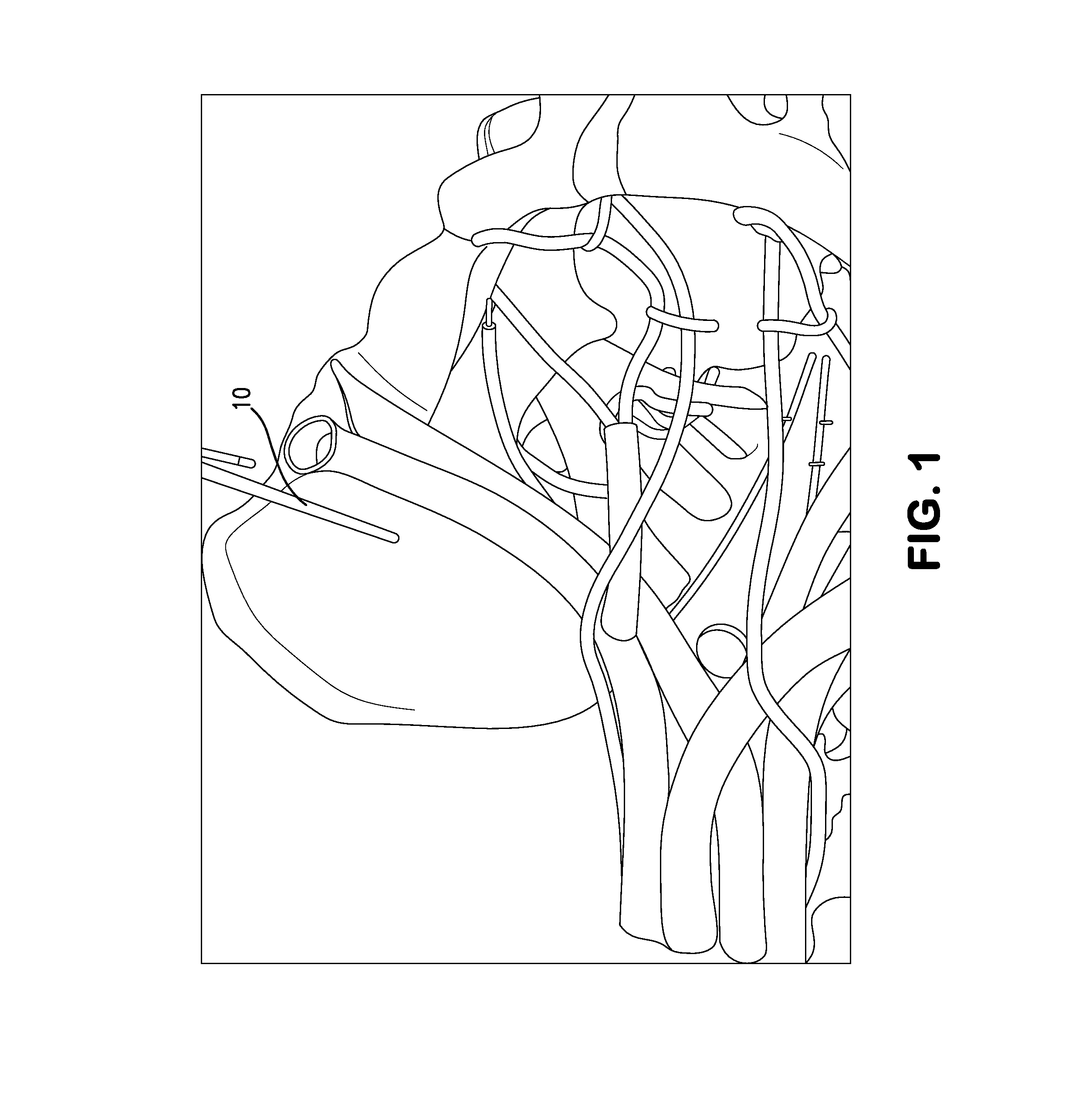
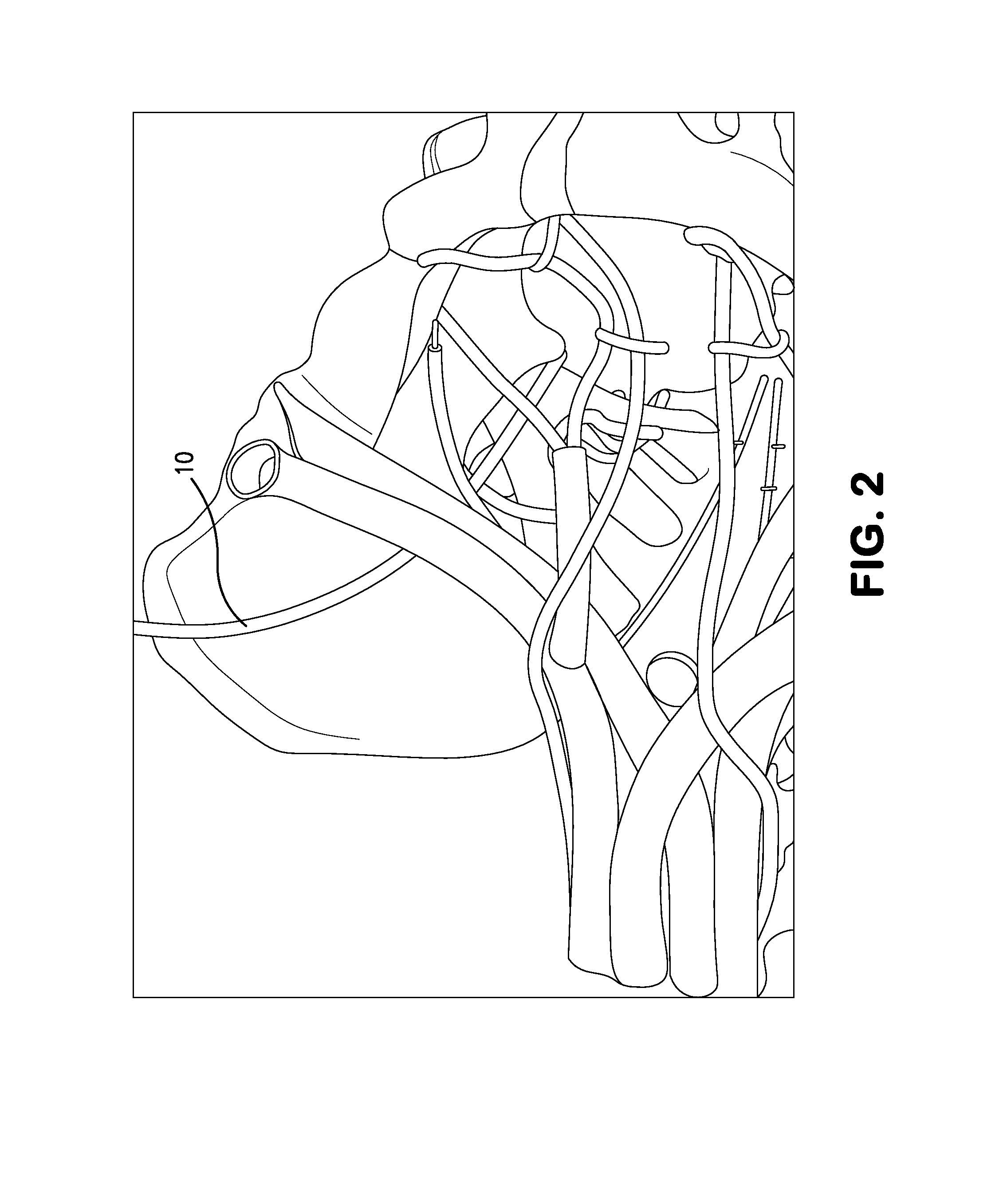
[0123]FIGS. 1-3 illustrate a model of the anatomical area of relevance to the present invention, with illustration of certain steps of the method of the present invention, while FIGS. 4-14 illustrate the tool and system of the present invention, all of which will be further described below.

[0124]I. System

A. The implant System

[0125]FIGS. 4-10 show an implant system for treating pelvic floor dysfunctions in humans.

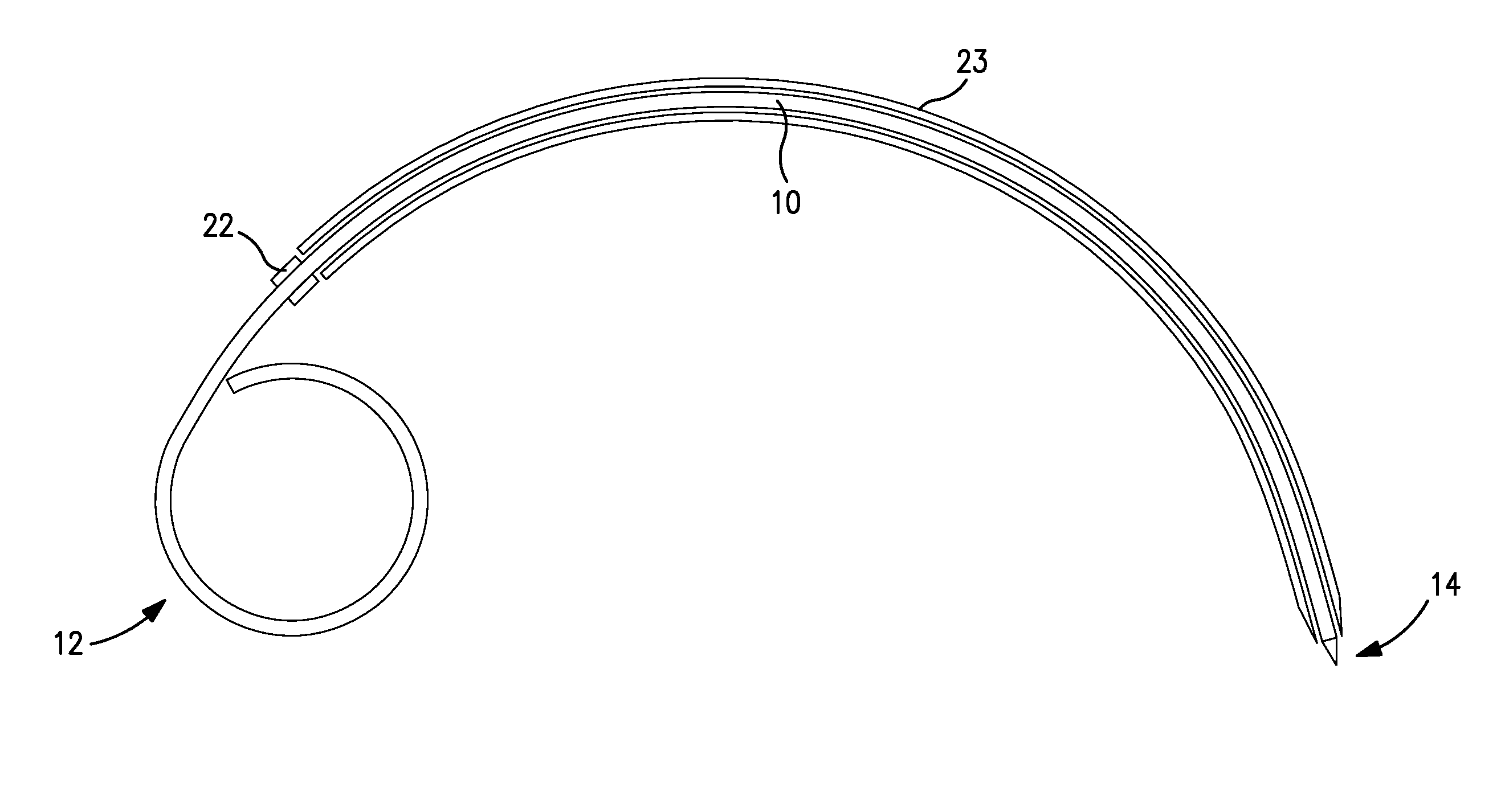
[0126]FIG. 4 shows a surgical system according to the invention which includes a curved tool 10 in a sleeve 23. Tool 10 has a handle portion 12 which can be curved or otherwise formed to be gripped by a surgeon or other user of the device. Tool 10 is further illustrated in FIG. 7. As shown, tool 10 can be formed from a cylindrical metal material and at one end forms curved grip portion 12 (segment A) and at the other end forms an engagement tip 14, which is formed at the end of a straight end or engagement segment 16 (segment 4, FIG. 7). Engagement tip 14 can be removable an...


PUM


Abstract

A collector electrode assembly which can be implanted by laparoscopy through the abdominal wall into the small pelvis of the human body includes a collector electrode for neurostimulation of nerves; a connection cable having an outer surface, said collector electrode being arranged at one end of said connection cable and comprising several outer segment electrodes which can be contacted individually and / or in groups and which are arranged axially one after another in the direction of the longitudinal extent of the collector electrode, wherein an insulating section is arranged axially between in each case two adjacent outer segment electrodes and permits electrical insulation of respective two adjacent outer segment electrodes; radially expandable fixing structures positioned on the collector electrode and radially expandable from a withdrawn position to a radially expanded position for fixing the collector electrode in place at said nerves; a visually perceptible direction marker on the outer surface of the connection cable, at least in a cable section which is spaced apart from axial ends of the connection cable and has an axial extent of at least 10 cm and / or at least 15% of total length of the connection cable, said direction marker indicating orientation of the connection cable to an operator using the assembly, and wherein the direction marker is designed and arranged in such a way that the identification of the orientation of the connection cable is possible at any desired axial section of the cable section having a maximum axial extent of 2 cm.

Description

BACKGROUND OF THE INVENTION[0001]The invention relates to a tool, system and method for treating at least one symptom of a pelvic floor and organ disorder and neuropathic pain by implanting a lead and electrode to the endopelvic portion of the pelvic nerves, nerves roots and / or plexuses using the tool, system and method according to the invention.[0002]Pelvic floor disorders adversely affect the health and quality of life of millions of people. Pelvic floor disorders include urinary control disorders such as urge incontinency, urge frequency, voiding efficiency, fecal control disorders, sexual dysfunctions, and pelvic pain.[0003]Lower urinary tract disorders affect the quality of life of millions of men and women over the world every year.[0004]Thirteen million Americans suffer from various types of urinary incontinence (UI). The most prevalent type of UI (22% of the total) is called Stress Incontinence (SUI). SUI is characterized by the unintended emission of urine during everyday ...

Claims


Application Information

Patent Timeline
IPC IPC(8): A61N1/05A61N1/36
CPCA61N1/0551A61N1/36107A61N1/36071A61N1/36007A61N1/0558
Inventor POSSOVER, MARC
Owner POSSOVER MARC
Learn More