
Engineering synthethic brain penetrating gene vectors

Active Publication Date: 2017-03-30
THE JOHN HOPKINS UNIV SCHOOL OF MEDICINE
View PDF1 Cites 1 Cited by
  • Summary
  • Abstract
  • Description
  • Claims
  • Application Information

AI Technical Summary

Benefits of technology

The patent describes a new way to deliver genes to brain cells using a special coating made of hydrophilic polymers. These coated genes penetrate the barriers in the brain and become active in target cells, resulting in highly effective gene expression. The technique uses a mixture of cationic polymers and a hydrophilic polymer called polyethylene glycol to create small particles called nanoparticles. These nanoparticles contain nucleic acid and a biodegradable polymer that can be designed to target specific brain cells. The nanoparticles are coated with polyethylene glycol to make them easily distributed throughout the brain. This technology can help develop new treatments for brain diseases.

Problems solved by technology

Patients with neurological diseases, including Parkinson's disease, Alzheimer's disease, brain tumors and most neurogenetic disorders, suffer from severe debilitating symptoms and lack of therapeutic options that provide curative treatment.
Viral gene vectors, though relatively efficient, have been limited by one or more drawbacks, including low packaging capacity, technical difficulties in scale-up, high cost of production [Thomas, et al., Nat Rev Genet, 2003.
However, non-viral gene vectors still face a number of barriers prior to reaching the target cells in the brain [O'Mahony, et al., J Pharm Sci, 2013.
This ‘brain tissue barrier’, regardless of administration method, hampers widespread distribution of macromolecules and nanoparticles in the brain, thereby limiting their coverage throughout the disseminated target area of neurological diseases [Voges, J., et al., Ann Neurol, 2003.
However, CED is unlikely to provide a significant benefit if particles remain entrapped in the brain parenchyma due to adhesive interactions and / or steric obstruction.
However, even following CED, the interactions between positively charged gene vectors and the negatively charged ECM, confine cationic nanoparticles to the point of injection and perivascular spaces, and limit their penetration into the brain parenchyma [MacKay, et al., Brain Res, 2005.

Method used


Image

  • Engineering synthethic brain penetrating gene vectors
  • Engineering synthethic brain penetrating gene vectors
  • Engineering synthethic brain penetrating gene vectors

Examples


example 1

on of Vectors to Shield the Positive Surface Charge Intrinsic to Cationic Polymer-Based Gene Vectors

[0181]Materials and Methods

Polymer Preparation

[0182]Methoxy PEG N-hydroxysuccinimide (mPEG-NHS, 5 kDa, Sigma-Aldrich, St. Louis, Mo.) was conjugated to 25 kDa branched polyethyleneimine (PEI) (Sigma-Aldrich, St. Louis, Mo.) to yield a PEG5k-PEI copolymer as previously described [30]. Briefly, PEI was dissolved in ultrapure distilled water, the pH was adjusted to 7.5-8.0 and mPEG-NHS was added to the PEI solution at various molar ratios and allowed to react overnight in 4° C. The polymer solution was extensively dialyzed (20,000 MWCO, Spectrum Laboratories, Inc., Rancho Dominguez, Calif.) against ultrapure distilled water and lyophilized. Nuclear magnetic resonance (NMR) was used to confirm a PEG:PEI ratio of 8, 26, 37 and 50. 1H NMR (500 MHz, D20): δ 2.48-3.20 (br, CH2CH2NH), 3.62-3.72 (br, CH2CH2O). The poly-L-lysine 30-mer (PLL) and PEG5K-PLL block copolymers were synthesized and ch...

example 2

or Particles are Stable Following Incubation in Physiological Environment

[0190]Materials and Methods

[0191]PEI nanoparticle stability was assessed by incubating nanoparticles in artificial cerebrospinal fluid (aCSF; Harvard Apparatus, Holliston, Mass.) at 37° C. and conducting dynamic light scattering every 30 mins for 24 hours. After 1 hour of incubation, a fraction of the nanoparticle solution was removed and imaged using TEM.

[0192]Results

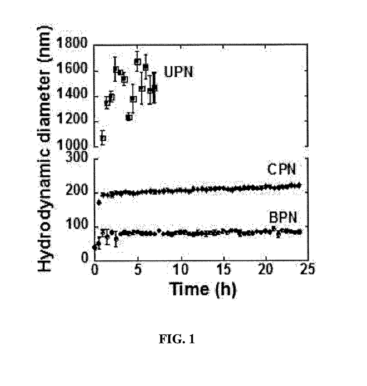
[0193]To predict the particle stability of gene vectors following in vivo administration, the in vitro stability in artificial cerebrospinal fluid (aCSF) was characterized over time at 37° C. (FIG. 1). UPN aggregated immediately after adding in aCSF. In 1 hour, the hydrodynamic diameter increased 8.3-fold and in 7 hours, the polydispersity was larger than 0.5, indicating loss of colloidal stability. CPN increased in diameter by 3-fold following incubation in aCSF and remained stable over 24 hours. BPN, formulated by the blend approach, exhibited i...

example 3

or Particles are Non-Toxic In Vitro and In Vivo

[0194]Materials and Methods

Cell Culture

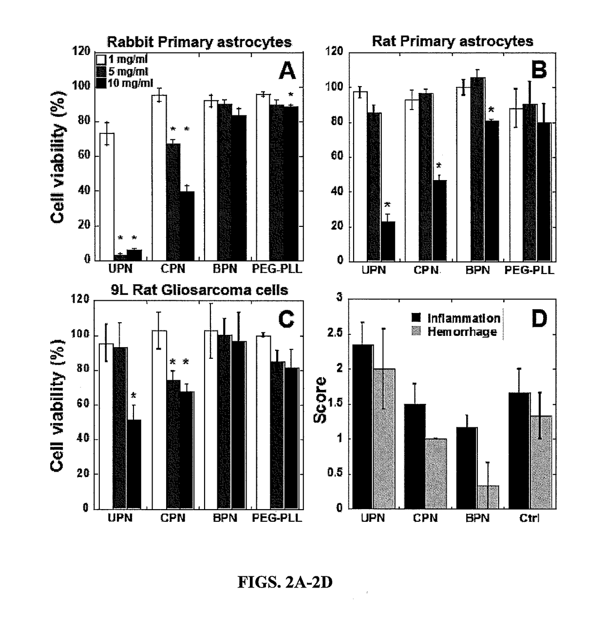
[0195]9 L gliosarcoma cells were provided by Dr. Henry Brem. 9 L immortalized cells were cultured in Dulbecco's modified Eagle's medium (DMEM, Invitrogen Corp., Carlsbad, Calif.) supplemented with 1% penicillin / streptomycin (pen / strep, Invitrogen Corp., Carlsbad, Calif.) and 10% heat inactivated fetal bovine serum (FBS, Invitrogen Corp., Carlsbad, Calif.). When cells were 70-80% confluent, they were reseeded in 96-well plates to assess toxicity and in 24-well plates to assess transfection and cell uptake of gene vectors. Rabbit primary astrocytes were provided by Dr. Sujatha Kannan. Mixed cell culture was prepared from day 1 neonatal rabbits and astrocytes were isolated using the conventional shake off method. Astrocytes were cultured in DMEM supplemented with 1% pen / strep and 10% FBS and passaged once; when cells were 70-80% confluent they were reseeded in 96-well plates for cell viability assay. ...


PUM

PropertyMeasurementUnit
Densityaaaaaaaaaa
Hydrophilicityaaaaaaaaaa
Biocompatibilityaaaaaaaaaa

Abstract

A synthetic gene delivery platform with a dense surface coating of hydrophilic and neutrally charged PEG, capable of rapid diffusion and widespread distribution in brain tissue, and highly effective gene delivery to target cells therein has been developed. Nanoparticles including nucleic acids, are formed of a blend of biocompatible hydrophilic cationic polymers and they hydrophilic cationic polymer conjugated to hydrophilic neutrally charged polymers such as polyethylene glycol. The nanoparticles are coated with polyethylene glycol at a density that imparts a near neutral charge and optimizes rapid diffusion through the brain parenchyma. Methods of treating a disease or disorder of the brain including administering a therapeutically effective amount of nano particles densely coated with polyethylene glycol are also provided.

Description

CROSS-REFERENCE TO RELATED APPLICATIONS[0001]This application claims priority to and the benefit of U.S. Ser. No. 61 / 991,898 filed May 12, 2014, U.S. Ser. No. 61 / 991,920 filed May 12, 2014 and U.S. Ser. No. 62 / 001,994 filed May 22, 2014, the disclosures of which are expressly incorporated hereby by reference.STATEMENT REGARDING FEDERALLY SPONSORED RESEARCH OR DEVELOPMENT[0002]This invention was made with Government Support under Agreement EB003558 and Agreement CA164789 awarded to Justin Hanes by the National Institutes of Health. The Government has certain rights in the invention.FIELD OF THE INVENTION[0003]This invention is generally in the field of gene delivery, and in particular, in delivering nucleic acids across biological barriers using coated particles to penetrate the brain parenchyma and achieve high level widespread transgene expression.BACKGROUND OF THE INVENTION[0004]Patients with neurological diseases, including Parkinson's disease, Alzheimer's disease, brain tumors a...

Claims


Application Information

Patent Timeline
IPC IPC(8): A61K48/00A61K9/00A61K9/51
CPCA61K48/0008A61K9/51A61K48/0075A61K9/5146A61K9/5192A61K9/5169A61K9/0085C12N15/88A61K47/60A61K47/6935
Inventor HANES, JUSTINSUK, JUNG SOOMASTORAKOS, PANAGIOTISWOODWORTH, GRAEMEZHANG, CLARK
Owner THE JOHN HOPKINS UNIV SCHOOL OF MEDICINE
Learn More