Optical physiologic sensors and methods

a physiologic sensor and optical technology, applied in the field of optical physiologic sensors and methods, can solve the problems of not providing the “bottom-line” tissue level insight that has long been missing, oxygen cannot be used, and cannot indicate whether there is enough, or too much, etc., to achieve rapid onset, long duration, and high-force body motions over short periods of time

Active Publication Date: 2017-04-27
REVEAL BIOSENSORS INC
View PDF2 Cites 17 Cited by
  • Summary
  • Abstract
  • Description
  • Claims
  • Application Information

AI Technical Summary

Benefits of technology

[0016]Additional aspects of the disclosure include physiologic sensors that are configurable to detect, by two-, or multi-wavelength photonic signal variation, the degree of physiologic loading due to physical exertion. Physical exertion can be roughly segmented into two categories: (1) anaerobic, which can provide rapid onset, high force body motions over short periods of time, such as weight lifting and sprinting, and (2) aerobic, which can support much longer duration, but relatively lower intensity continuous body motions, such as marathon running. Athletes training for, and performing in one or the other type of activity have been found to benefit from narrowly defined, performance-specific types of training and conditioning exercise sessions. Examples of this include anaerobic-type performance needing multiple brief, high force generation cycles, with full recovery between, which has been found to stimulate up-regulation of enzyme systems needed for generation of adenosine triphosphate (ATP) by glycolysis. On the other hand, long distance runners have been found to benefit most from prolonged, fully aerobic exercise sessions, avoiding even brief periods of anaerobic metabolism, to up-regulate their enzyme systems needed for converting fats for use in aerobic energy conversion. Until the discovery of the PI signal, there has not been a convenient, objective, wearable method of sensing when the athlete is using primarily anaerobic, vs. aerobic energy conversion chemistry during exercise.
[0017]Another embodiment may integrate both PI and reflectance SpO2 sensor functions, where these two biometrics each provide highly relevant, related, and complementary information in many useful applications. In this integrated format, such as for use on the chest and abdomen of a premature infant, the SpO2 monitor function primarily provides an indication of the adequacy of the lungs to obtain oxygen, and the computed PI value provides an index of skin tissue oxygenation, as a surrogate for oxygen delivery by the blood to the brain and other vital internal organs. Existing SpO2 monitors are well known to be easily compromised by sensor motion-induced optical signal artifacts that temporarily prevent accurate computation of the SpO2 value, which, in turn, may trigger false alarms and, alternatively, may result in failure to initiate an alarm during real alarm conditions. On the other hand, the PI sensor is more quickly responsive to changes in oxygen delivery at the skin tissue level, and is inherently immune to sensor motion-induced signal artifact due to the tandem pattern of the variations that occur in the raw signal values with sensor motion. Thus, the PI value can be safely and effectively relied upon to provide a backup index to eliminate false SpO2 alarms and to avoid missing real SpO2 alarm conditions accompanied by sensor motion.

Problems solved by technology

While each of these separate measurements are known to provide significant information, this currently available data, even with expert analysis, cannot provide the “bottom-line” tissue-level insight that has long been missing.
The recently introduced Lumee™ sensor (Profusa), measures the oxygen level in tissue, but cannot indicate whether there is enough, or too much, oxygen.
Also, the new sweat lactate sensor from Kenzen indicates when the skin is anaerobic, but cannot work when there is no sweat production and does not sense when too much oxygen is present.
Without accurate, objective tissue-level information, the limitations of blood and tissue oxygen metrics, human sensory misperceptions and subjective clinician assessment errors in this critical part of medical care can inadvertently result in permanent vital organ tissue damage and possibly death.
While “medical device-like” vital sign monitoring devices have recently been re-packaged into consumer-friendly “activity tracker” devices, the same limitations remain with respect to their delivery of physiologic information.

Method used


Image

  • Optical physiologic sensors and methods
  • Optical physiologic sensors and methods
  • Optical physiologic sensors and methods

Examples


examples

[0092]Sepsis

[0093]Another example of an application of the disclosed technology is directed to an alternative process for continuous re-evaluation and adjustment of the PI zero. Many potentially useful applications of the disclosed apparatus and methods will start when the monitored subject is not in a healthy, resting state. For example, at the initial presentation of a patient with early sepsis to a hospital emergency department, there is likely already a significant pathologic decrease of the blood perfusion to the skin, resulting in the skin tissue status being within the person's previous anaerobic range; i.e. negative PI value. Simply using the presenting abnormal physiologic conditions to set the PI zero will result in an indeterminate negative deviation of the PI zero relative to that person's previous healthy, resting PI zero. As effective treatment is administered, and the patient's physiologic status improves back toward normal health, the transition between negative PI a...


PUM


Abstract

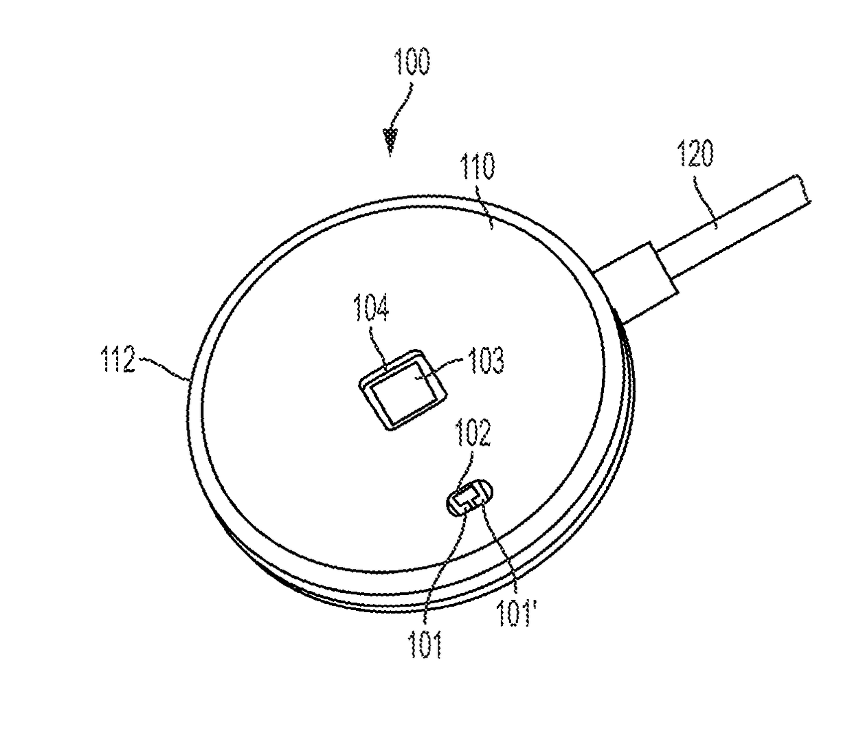
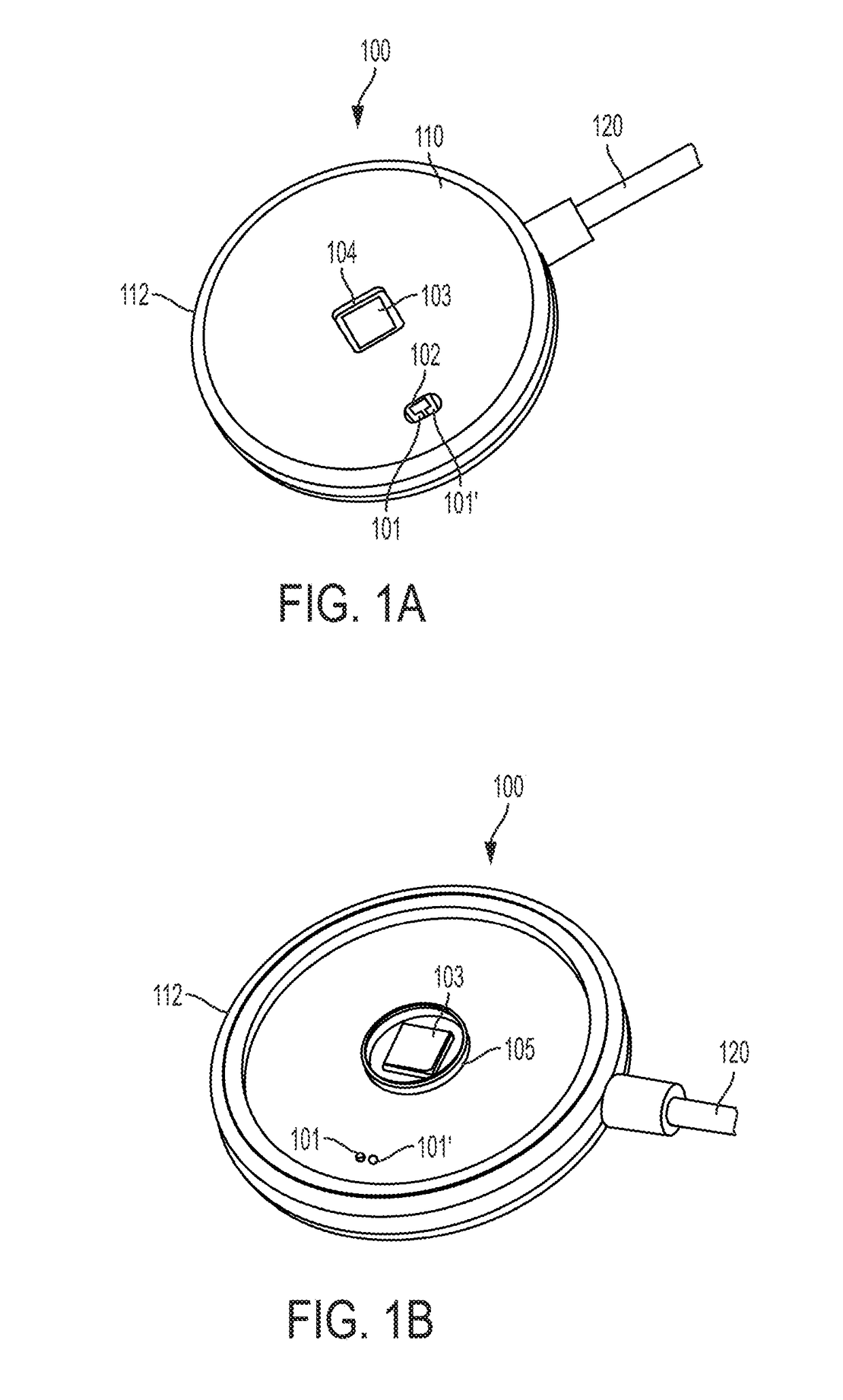
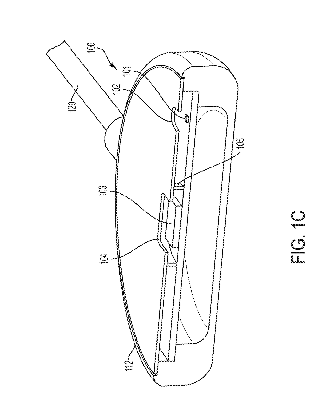
Physiologic sensors and methods of application are described. These sensors function by detecting recently discovered variations in the spectral optical density at two or more wavelengths of light diffused through the skin. These variations in spectral optical density have been found to consistently and uniquely relate to changes in the availability of oxygen in the skin tissue, relative to the skin tissue's current need for oxygen, which we have termed Physiology Index (PI). Current use of blood gas analysis and pulse oximetry provides physiologic insight only to blood oxygen content and cannot detect the status of energy conversion metabolism at the tissue level. By contrast, the PI signal uniquely portrays when the skin tissue is receiving ‘less than enough oxygen,’‘just the right amount of oxygen,’ or ‘more than enough oxygen’ to enable aerobic energy conversion metabolism. The PI sensor detects one pattern of photonic response to insufficient skin tissue oxygen, or tissue hypoxia, (producing negative PI values) and a directly opposite photonic response to excess tissue oxygen, or tissue hyperoxia, (producing positive PI values), with a neutral zone in between (centered at PI zero). Additionally, unique patterns of PI signal response have been observed relative to the level of physical exertion, typically with a secondary positive-going response trend in the PI values that appears to correspond with increasing fatigue. The PI sensor illuminates the skin with alternating pulses of selected wavelengths of red and infrared LED light, then detects the respective amount of light that has diffused through the skin to an aperture located a lateral distance from the light source aperture. Additional structural features include means of internally excluding light from directly traveling from the light emitters to the photodetector within the sensor. This physiology sensor and methods of use offer continuous, previously unavailable information relating to tissue-level energy conversion metabolism. Several alternative embodiments are described, including those that would be useful in medical care, athletics, and personal health maintenance applications.

Description

CROSS-REFERENCE[0001]This application claims the benefit of U.S. Provisional Application No. 62 / 246,374, filed Oct. 26, 2015, entitled OPTICAL PHYSIOLOGIC SENSORS AND METHODS which application is incorporated herein by reference.BACKGROUND[0002]One of the primary life-dependencies of humans is continuous delivery of oxygen by the lungs and blood circulatory system to all tissues of the body in sufficient quantity to maintain aerobic metabolism; thus avoiding tissue injury from too little, or too much oxygen. To approximate this information, clinicians must mentally correlate measurements of breathing gas oxygen fraction, breathing rate, heart rate, cardiac output, blood hemoglobin level, arterial blood hemoglobin-oxygen saturation (“blood gas,” or SaO2), and pulse oximetry (SpO2), along with a subjective evaluation of arterial blood flow distribution. While each of these separate measurements are known to provide significant information, this currently available data, even with expe...

Claims


Application Information

Patent Timeline
Patent Type & Authority Applications(United States)
IPC IPC(8): A61B5/1455A61B5/00
CPCA61B5/14552A61B5/6823A61B5/0022A61B5/0004A61B5/6824A61B5/6833A61B5/0075A61B2562/185
Inventor HATCH, GUY MEREDITH
Owner REVEAL BIOSENSORS INC
Why Eureka
Try Eureka