Pharmaceutical formulations that form gel in situ

a technology of pharmaceutical formulations and gels, applied in the field of ophthalmic disorders, can solve the problems of inability to accurately diagnose infectious conjunctivitis, inability to cure infections, etc., to achieve the effect of prolonging the residence time of the vehicle, and reducing the risk of infection

Active Publication Date: 2017-09-21
IVIEW THERAPEUTICS
View PDF1 Cites 7 Cited by
  • Summary
  • Abstract
  • Description
  • Claims
  • Application Information

AI Technical Summary

Benefits of technology

[0004]Ophthalmic topical drug delivery is one of the important methods of application, but the existence of cornea barrier, tear dilution and lacrimal passage drainage effect limit the treatments and bioavailability of many topical ophthalmic preparations. The conventional liquid ocular formulation is eliminated from the precorneal area immediately upon instillation because of lachrymation and effective nasolacrimal drainage. See, e.g., VHL Lee et al., J. Pharm. Sci., 1979; 68: 673-84. Various preparations, such as ointments, suspensions, inserts, and hydrogels, have been develo

Problems solved by technology

Because antibiotic drops are ineffective against viral conjunctivitis, treatments for such infections can only relieve symptoms.
Diagnosis of the various causative agents, such as bacteria, virus, or fungus, in infectious conjunctivitis is not economically feasible because accurate diagnosis requires sophisticated laboratory culture not easily integrated into the average healthcare practice.
Because accurate diagnosis is impractical, most conjunctivitis is presumed to be bacterial without culturing and is treated with antibiotics.
Antibiotic treatment is suboptimal because it is ineffective against viral or fungal conjunctivitis.
Ophthalmic topical drug delivery is one of the important methods of application, but the existence of cornea barrier, tear dilution and lacrimal passage drainage effect limit the treatments and bioavailability of many topical ophthalmic preparations.
However, they have not been used extensively because of some drawbacks, such as blurred vision with ointments or low patient compliance with inserts.
However, at a low concentration, PVP-I will degrade quickly, and its concentration cannot be effectively maintained during storage or at the infected site due to the tear barrier effects.
However, as a result of strong oxidizing potential and acidity of water-soluble PVP-I polymer material,

Method used


Image

  • Pharmaceutical formulations that form gel in situ
  • Pharmaceutical formulations that form gel in situ
  • Pharmaceutical formulations that form gel in situ

Examples


example 1

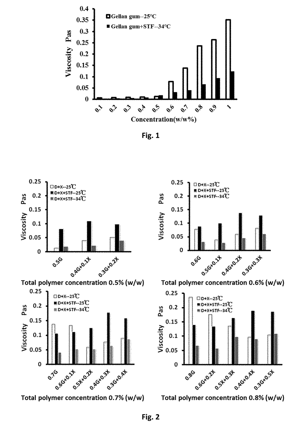
[0062]Preparation of solution of deacetylated gellan gum (DGG) (Kelcogel-Cg-La gellan gum, food grade gellan gum, CAS: 71010-52-1: E418, particle size: ˜42 mesh (355 μm), purchased from CPKelco): DGG was dissolved in deionized water and the solution was stirred in an 80° C. water bath for 1 hour, cooled to the room temperature, allowed to stand until the material is fully swollen, and used to prepared solutions of 0.1% to 1.0% (w / w) concentrations.

[0063]Preparation of simulated tear fluid (STF): NaHCO3 2.18 g; NaCl 6.78 g; CaCl2.2H2O 0.084 g; KCl 1.38 g; dissolve in 1000 mL deionized water: DGG solution and simulated tear fluid were mixed at the 40:7 ratio, and the viscosity of the DGG solution was measured before and after mixing with stimulated tear fluid with a rotary rheometer at 25° C. and at 34° C., respectively. The viscosity change was shown in FIG. 1. For the DGG solution in a concentration range of 0.1% to 1.0% (w / w), its viscosity was reduced significantly under simulated...

example 2

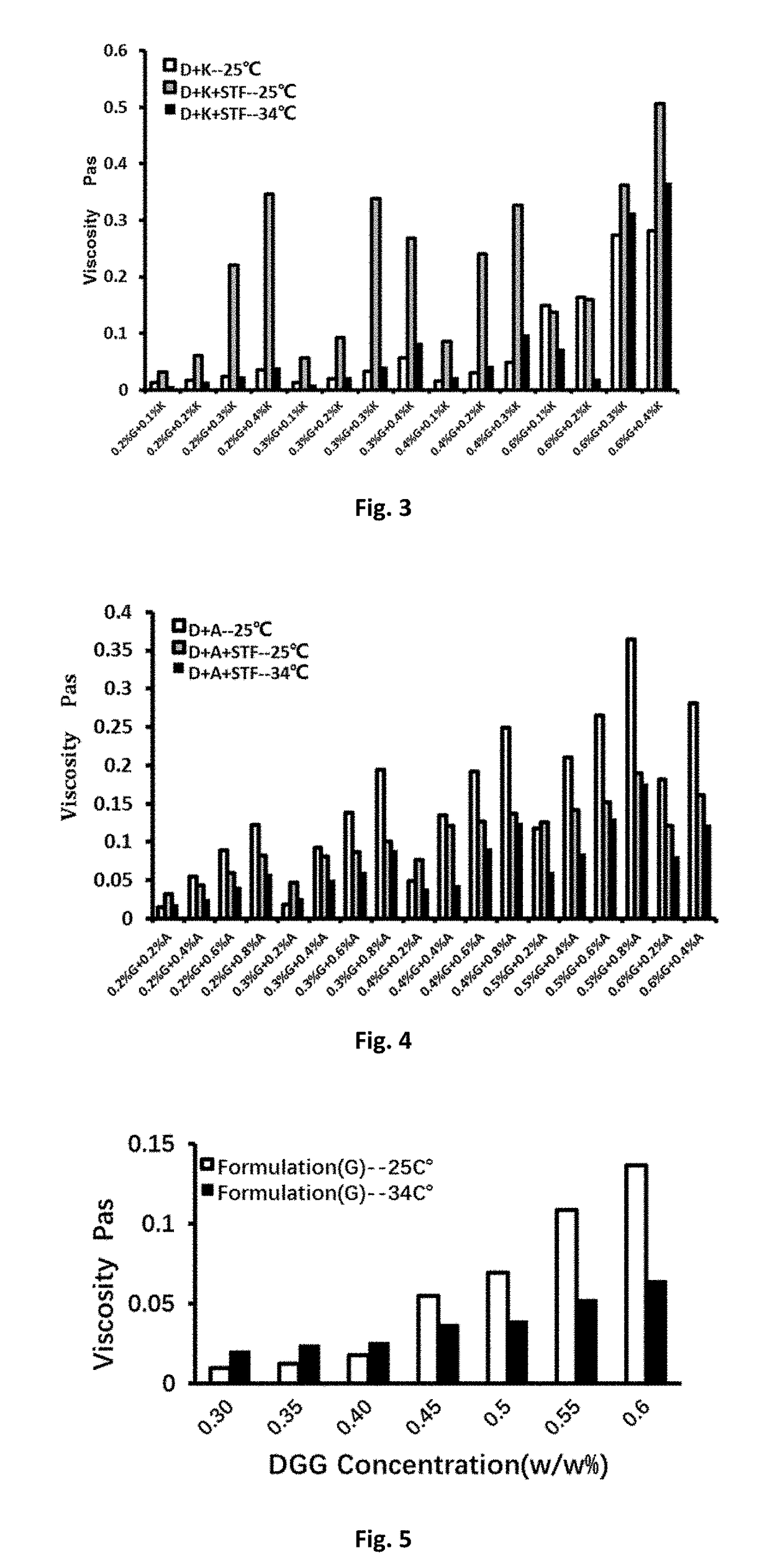
[0064]DGG-Xanthan mixed solution: DGG and xanthan were weighed and used at a certain proportion and added into deionized water. The mixture was stirred in an 80° C. water bath for 1 hour after the dispersion of DGG and xanthan in the water, cooled to the room temperature, and allowed to stand until fully swollen. The morphological scoring of the deacetylated gellan gum-xanthan mixed solution before and after adding simulated tear fluid was evaluated according to the following criteria: (1) thin liquid: 1-3 points; (2) thick gelatinous form: 4-6 points; (3) gel state: 7-9 points.

TABLE 1Morphological scoring of the DGG-xanthan solutions beforeand after adding STF.ScoringScoringDGGXanthanScoringD + X + STFD + X + STF(%, w / w)(%, w / w)D + X25° C.(25° C.)34° C. Δ0.30.11210.21320.32310.43520.53630.63740.40.12310.22310.32420.45610.50.12310.22420.33410.602420.13300.24400.35500.4550

[0065]As shown in Table 1 and FIG. 2, the viscosity change tendency of DGG-xanthan solution was consistent with t...

example 3

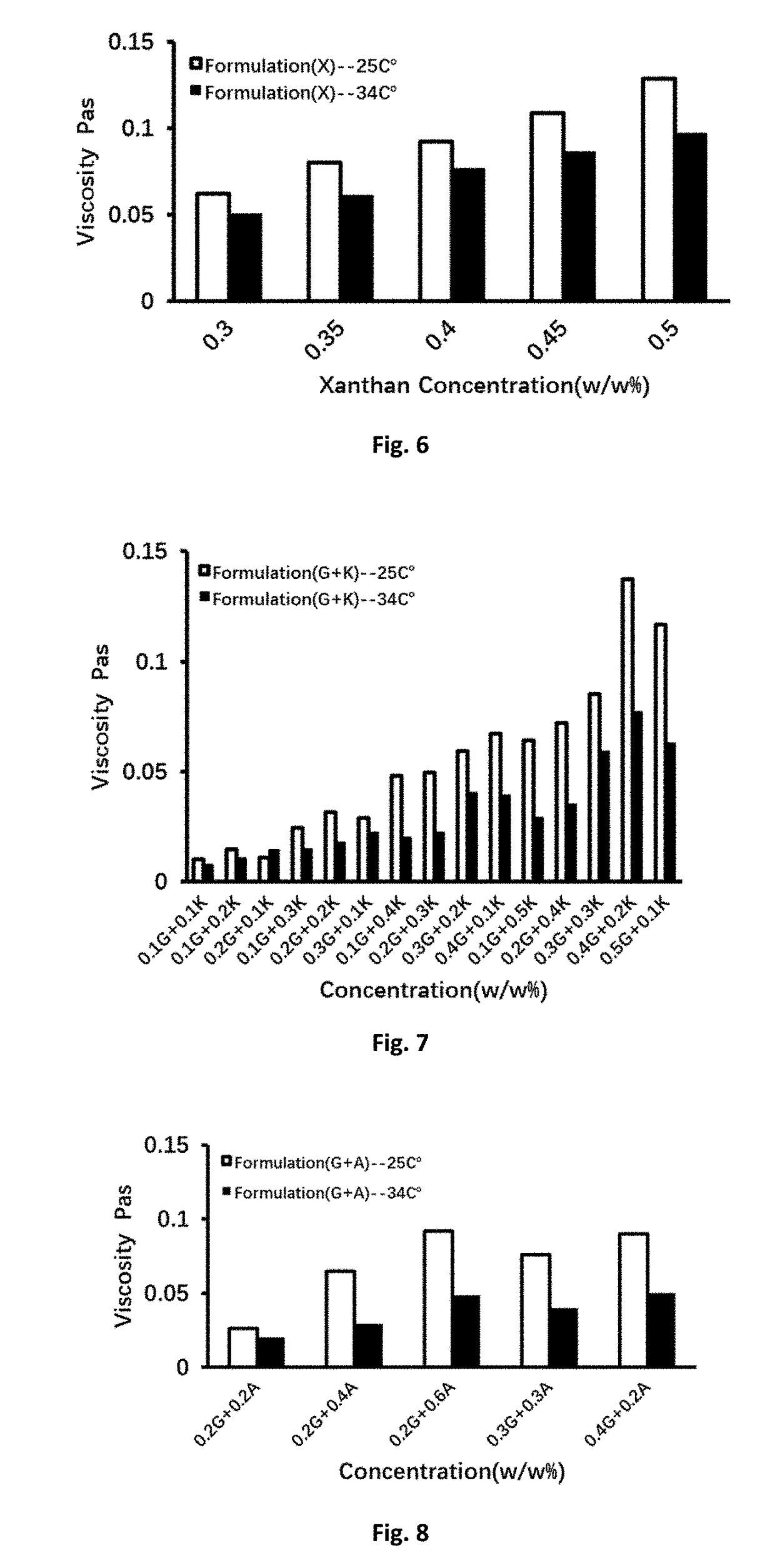
[0066]DGG-Kappa-Carrageenan compound solution: DGG and carrageenan were weighed and used at a certain proportion, added into deionized water, and the mixture was slowly stirring in an 80° C. water bath for 1 hour after being well-dispersed. It was then cooled to the room temperature and allowed to stand until fully swollen. The morphological scoring of the DGG-kappa-carrageenan mixed solutions before and after adding tear fluid was evaluated according to the above-mentioned criteria.

TABLE 2The morphological scoring of the DGG-kappa-carrageenanmixed solution before and after adding STF.Kappa-DGGCarrageenan25° C.-D + K + STF(%, w / w)(%, w / w)D + KD + K + STF34° C. Δ0.20.11100.21100.32200.42530.30.11100.21210.32750.43850.40.12200.22640.33630.50.13300.60.14620.24620.37810.4891

[0067]As the result shown in Table 2 and FIG. 3, the viscosity change tendency of DGG-carrageenan mixed was consistent with DGG solution alone at both the room temperature 25° C. and simulated physiological condition...


PUM

PropertyMeasurementUnit
Fractionaaaaaaaaaa
Fractionaaaaaaaaaa
Fractionaaaaaaaaaa

Abstract

The present invention provides aqueous formulations containing an anti-infection agent, a biocompatible polysaccharide, an osmotic pressure regulator, a pH regulator, and water, wherein a gel containing the therapeutic agent is formed in situ upon instillation of the formulations onto the skin and a body cavity of a subject. The formulations of this invention are useful for treating infectious diseases of skin or a body cavity (e.g., eye, nose, or vagina) of a subject.

Description

CROSS-REFERENCE TO RELATED APPLICATION[0001]This application claims priority to U.S. Application No. 62 / 246,073, filed on Oct. 25, 2015, the contents of which are incorporated herein in their entirety.BACKGROUND OF THE INVENTION[0002]Infectious conjunctivitis is an ophthalmic disorder characterized by inflammation of the conjunctiva secondary to invasion of a microbe. Microbes capable of causing conjunctivitis in humans include bacteria (including Mycobacteria sp), viruses, fungi, and amoebae. Current treatments for bacterial conjunctivitis consist of antibiotic drops. Because antibiotic drops are ineffective against viral conjunctivitis, treatments for such infections can only relieve symptoms. Treatments for fungi and amoeba conjunctivitis consist of a small selection of medications which lack sufficient anti-bacterial or anti-viral activity and are sometimes toxic to the ocular surface.[0003]Diagnosis of the various causative agents, such as bacteria, virus, or fungus, in infecti...

Claims


Application Information

Patent Timeline
IPC IPC(8): A61K47/36A61K31/155A61K9/00A61K31/79A61K9/06A61K47/02
CPCA61K47/36A61K9/06A61K31/155A61K9/0048A61K31/79A61K47/02A61K47/10A61K47/26A61K47/32A61K33/18A61K9/0014A61P27/02A61P31/00
Inventor BALDWIN, JOHN J.WEI, GANG
Owner IVIEW THERAPEUTICS
Why Eureka
Try Eureka