Three-dimensional bioprinted artificial cornea

a bioprinting and artificial cornea technology, applied in the field of artificial cornea, can solve the problems of long wait lists, limited supply of donor tissue, and even more difficult access to donor tissue, and achieve the effect of changing the clinical landscape and restoring vision

Inactive Publication Date: 2017-10-05
RGT UNIV OF CALIFORNIA
View PDF2 Cites 10 Cited by
  • Summary
  • Abstract
  • Description
  • Claims
  • Application Information

AI Technical Summary

Benefits of technology

[0014]By integrating the emerging technologies in the multidisciplinary domains of nanotechnology, 3D bioprinting, and regenerative medicine, we have developed artificial corneas to change the clinical landscape by eliminating the current dependency on corneal donor tissue and

Problems solved by technology

Disease or damage to one or more layers of the cornea can lead to blindness that is commonly treated by corneal transplant.
Although the surgery has a high success rate, the supply of donor tissue is limited, and wait lists can be long.
In the developing world, access to donor tissue is even more difficult.
Further, while human donor transplants are the standard treatment for corneal blindness, the complications and limitations inherent in them have prompted development of synthetic corneal substitutes.
Although Kpros have been available for many years in various forms, the fabrication of synthetic stromal equivalents with the transparency, biomechanics, and regenerative capacity of human donor corneas remain a formidable challenge.
Further, the

Method used


Image

  • Three-dimensional bioprinted artificial cornea
  • Three-dimensional bioprinted artificial cornea
  • Three-dimensional bioprinted artificial cornea

Examples


example 1

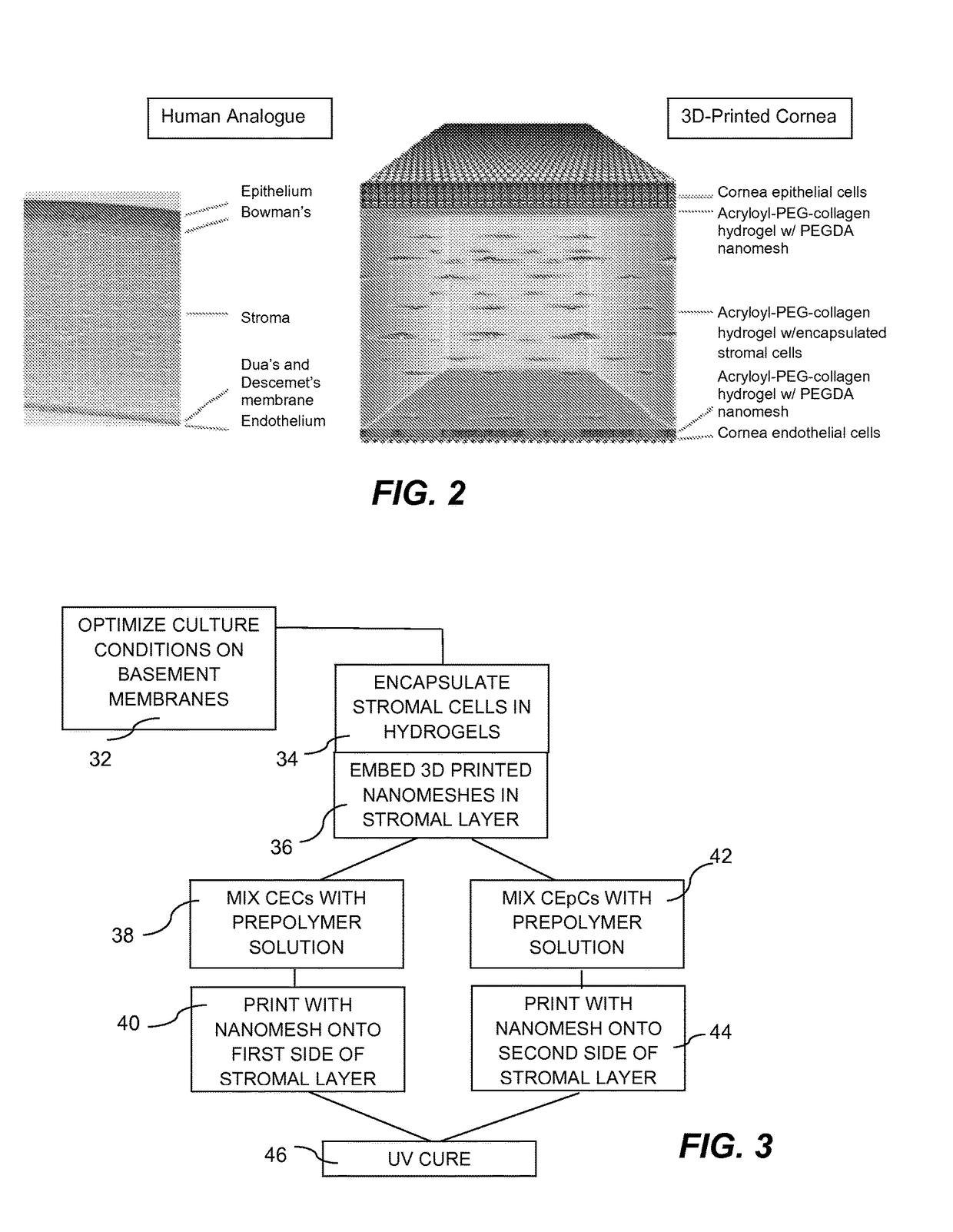
EpCs, CECs, and Stromal Cells on a Basement Membrane

[0035]Cornea epithelial cells (CECs) undergo continuous renewal from limbal stem or progenitor cells (LSCs), and deficiency in LSCs or corneal epithelium, which turns cornea into a non-transparent, keratinized skin-like epithelium, causes corneal surface disease that leads to blindness. How LSCs are maintained and differentiated into corneal epithelium in healthy individuals, and which molecular events are defective in patients have been largely unknown.

[0036]Traditionally, the LSC growth and expansion process requires mouse 3T3 feeder cells, which carry the risk of contamination from animal products, thereby rendering it unsuitable for creating clinically-viable 3D bioprinted corneas. To overcome these obstacles, an in vitro feeder-cell-free, chemically-defined cell culture system to grow LSCs from rabbit and human donors, was developed to enable generation and expansion of a homogeneous population of LSCs, and subsequent differen...

example 2

nting

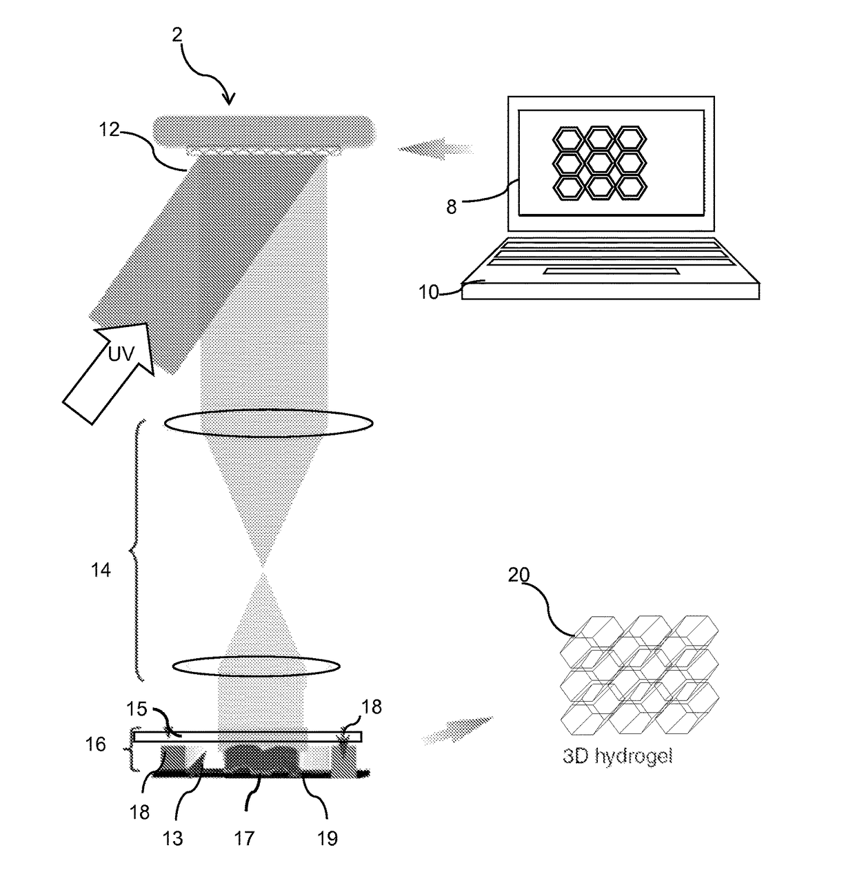
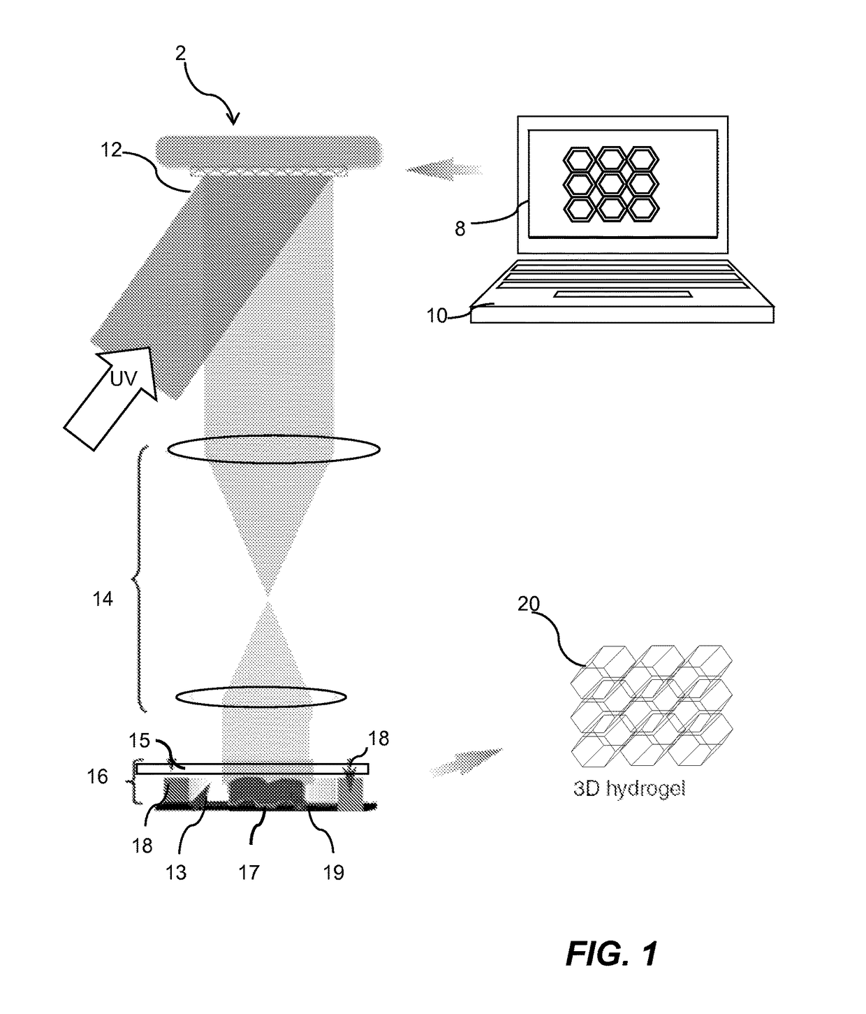
[0042]The 3D bioprinting platform offers a rapid biofabrication approach for constructing cell-laden hydrogel scaffolds that 1) have complex user-defined 3D geometries composed of a naturally derived biomaterial; 2) allow for consistent 3D distribution of cells encapsulated within the hydrogel; 3) support cell viability and proliferation; and 4) feature dynamic, multi-scale mechanical cell-scaffold interactions. Importantly, these constructs enable control and integration of complex 3D geometries while providing a physiologically-relevant internal 3D distribution of encapsulated cells. Through such precise control of spatial and temporal distributions of biological factors in 3D scaffolds, we are able to evaluate the interactions of cells with extracellular matrix (ECM) proteins at the nanometer length scale, with the ultimate goal of creating advanced, clinically translatable biomimetic scaffolds.

[0043]Using 3D bioprinting, artificial corneas are fabricated using the same dime...

example 3

als for Cornea Tissues

[0045]Collagen has been used extensively as a biomaterial for corneal tissue engineering, as it comprises the main component of corneal extracellular matrix (ECM). Collagen, as a matrix constituent, has been demonstrated to support epithelial cells in forming a protective layer and to promote re-innervation by neurons. A chemically-crosslinked biosynthetic collagen matrix has shown significant promise in a phase I clinical trial. In order to modulate the degradation and mechanical properties of a collagen matrix, most studies have used chemical crosslinking approaches, which are largely incompatible with cell encapsulation. Acryloyl-PEG-collagen (Ac-Col) offers an excellent alternative for corneal tissue engineering due to its biocompatibility, optical properties, and ability for photopolymerization. Preliminary tests have been performed to assess the optical properties of a stromal cell-laden film made of GelMA, which is an Ac-Col analogue. FIG. 6 illustrates ...


PUM


Abstract

An artificial cornea is fabricated by separately culturing live stromal cells, live corneal endothelial cells (CECs) and live corneal epithelial cells (CEpCs), and 3D bioprinting separate stromal, CEC and CEpC layers to encapsulate the cells into separate hydrogel nanomeshes. The CEC layer is attached to a first side of the stromal layer and the CEpC layer to a second side of the stromal layer to define the artificial cornea.

Description

RELATED APPLICATIONS[0001]This application claims the benefit of the priority of U.S. Provisional Application No. 62 / 054,924, filed Sep. 24, 2014, which is incorporated herein by reference in its entirety.FIELD OF THE INVENTION[0002]The invention relates to 3D bioprinting of artificial tissue and more specifically to an artificial cornea produced using 3D bioprinting.BACKGROUND OF THE INVENTION[0003]Disease or damage to one or more layers of the cornea can lead to blindness that is commonly treated by corneal transplant. Approximately 40,000 patients undergo corneal transplant surgery in the United States every year. The vast majority of these people receive a replacement cornea from a human donor. Although the surgery has a high success rate, the supply of donor tissue is limited, and wait lists can be long. In the developing world, access to donor tissue is even more difficult. Further, while human donor transplants are the standard treatment for corneal blindness, the complicatio...

Claims


Application Information

Patent Timeline
IPC IPC(8): A61L27/38B33Y80/00C12N5/079B33Y10/00A61L27/52A61L27/48
CPCA61L27/3891A61L27/3808A61L27/3813A61L27/3834A61L27/52A61L27/48C12N2513/00B33Y10/00B33Y80/00A61L2430/16A61L2400/12C12N2533/54C12N2533/30C12N5/0621C12M33/00A61L27/50
Inventor ZHANG, KANGCHEN, SHAOCHENQU, XINOUYANG, HONG
Owner RGT UNIV OF CALIFORNIA
Why Eureka
Try Eureka