

Shear controlled release for stenotic lesions and thrombolytic therapies
a stenotic lesions and controlled release technology, applied in the direction of drug compositions, peptide/protein ingredients, extracellular fluid disorders, etc., can solve the problems of unproposed targeting strategies based on such parameters, and alternative approaches have not been suggested or developed for drug delivery
- Summary
- Abstract
- Description
- Claims
- Application Information
AI Technical Summary
Benefits of technology
Problems solved by technology
Method used
Image

Examples
example 1
[0434]PEG-PLGA (50:50 MW 4:17 kDA) or PLGA (50:50 MW 17 kDa) nanoparticles were prepared as follows. The COOH-PEG-PLGA or COOH-PLGA polymer was conjugated with the binding peptides (CREKA (SEQ ID NO: 1) or CRKRLDRNK (SEQ ID NO: 2)) using a maleimide-PEG-NH2 linker. First, the cysteine ends of the peptides were conjugated to the maleimide end of the PEG linker in PBS buffer, at pH 6.5. Second, carboxyl functionalized PEG-PLGA polymer was pre-activated with 1-ethyl-3-(3-dimethylaminopropyl) (EDC) and N-hydroxysuccinimide (NETS) and subsequently reacted with the corresponding PEG modified peptides in PBS / DMSO mixture at room temperature for about 4 hours. The final products were purified by dialysis and a lyophilized powder was obtained. The conjugation reaction was confirmed by 1E1 NMR. The peptide conjugated polymer was then dissolved with coumarin a hydrophobic dye either in ethyl acetate or DMSO and the nano particles were prepared by w / o / w emulsion or simple solvent displacement m...
example 2
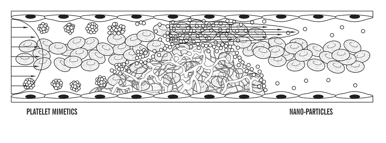
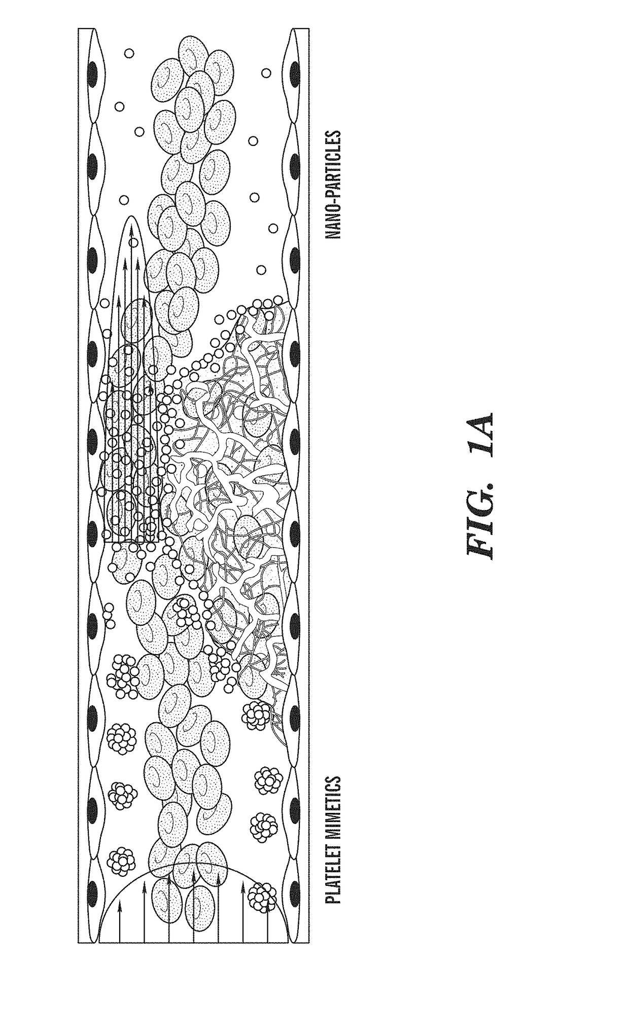
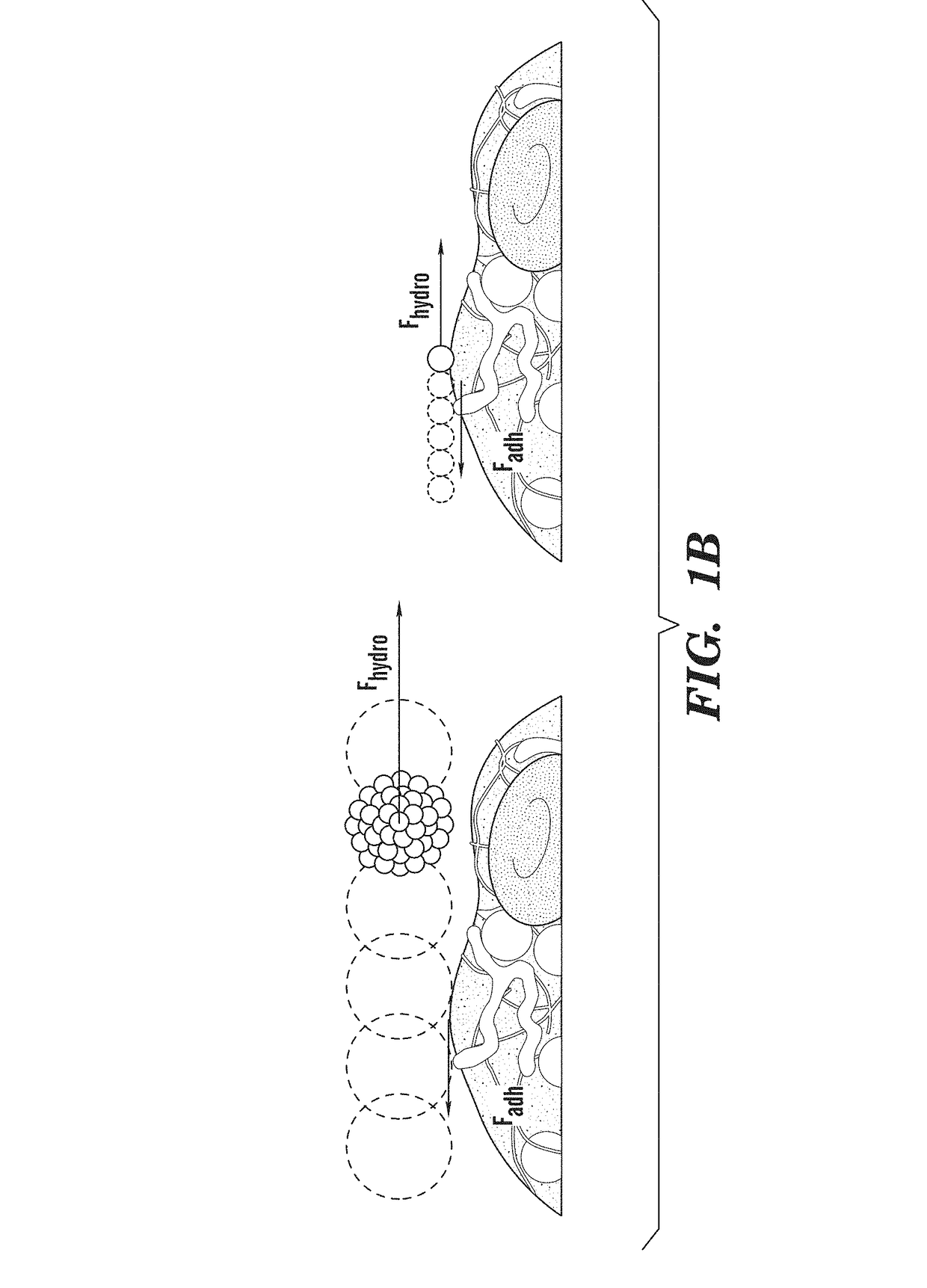
ivated Platelet Mimetics for Drug Targeting to Obstructed Blood Vessels
Materials and Methods
[0439]Nanoparticle Preparation:
[0440]Nanoparticles (NPs) were prepared from PLGA (50:50, 17 kDa, acid terminated; Lakeshore Biomaterials, AL) using a simple solvent displacement method (C. E. Astete &C. M. Sabliov, Journal of Biomaterials Science, Polymer Edition 17, 247 (2006)). The fluorescent hydrophobic dye, coumarin, was included in the NPs to enable visualization and quantitation in this study. Briefly, 1 mg / ml of polymer was dissolved with coumarin in dimethyl sulfoxide (DMSO, Sigma, MO), dialyzed against water at room temperature, and the nanoparticles were allowed to form by solvent displacement. The size distribution and morphology of the formed NPs were characterized using Dynamic Light Scattering (DLS), Scanning Electron Microscopy (SEM) and Transmission Electron Microscopy (TEM).
[0441]Fabrication of Platelet Mimetics—
[0442]The PLGA NPs were centrifuged and concentrated to a 10 mg...
example 3
ess Controlled Release from RBCs
[0483]Red blood cells ghosts were prepared using hypotonic hemolysis method. In brief, RBC were centrifuged from blood (2000 g, 10 min) and resuspended in calcium / magnesium free diluted PBS (PBS to DD water vol ratio of 1:10). The cells were allowed to incubate for 15 minutes at 4° C. and then centrifuged (12,000 g, 10 min). This process was repeated four times. Afterwards the cells were loaded with FITC-dextran by incubating the cells with 5 mg / ml dextran in diluted PBS for 1 hour at 4° C. The cells were centrifuges, suspended in PBS buffer with Ca / Mg and allowed to reseal in a 37° C. incubator for more than 2 hr. Following the resealing procedure the cells were washed in PBS for four times to remove any residuals in solution. FIG. 8 shows a fluorescence image of RBC ghosts loaded with FITC-dextran taken five days after preparation of FITC-dextran loaded ghosts.
[0484]A suspension of FITC-dextran loaded RBC ghosts was infused through a device without ...
PUM
 Abstract
Description
Claims
Application Information