

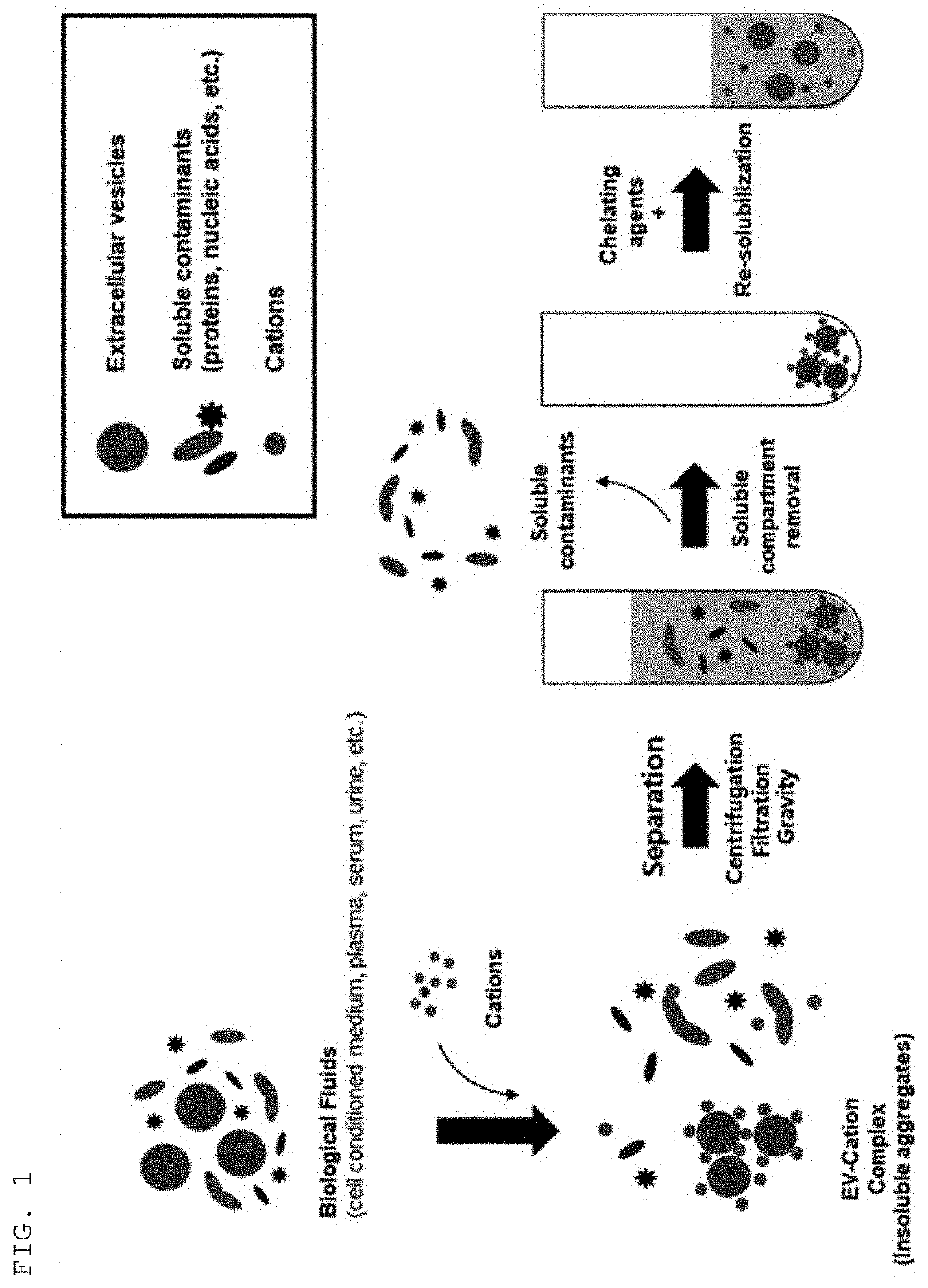
Method for isolating extracellular vesicles using cations
a technology of extracellular vesicles and cations, which is applied in the field of isolating extracellular vesicles using cations, can solve the problems of difficult to develop antibodies and protein ligands at high efficiency, limited examples of materials having such selective binding properties, and difficult to isolate extracellular vesicles efficiently. , to achieve the effect of maintaining the morphology or properties of extracellular vesicles, preventing the exposure of samples, and efficiently
- Summary
- Abstract
- Description
- Claims
- Application Information
AI Technical Summary
Benefits of technology
Problems solved by technology
Method used
Image

Examples
example 1
Purification and Analysis of Sample Extracellular Vesicles
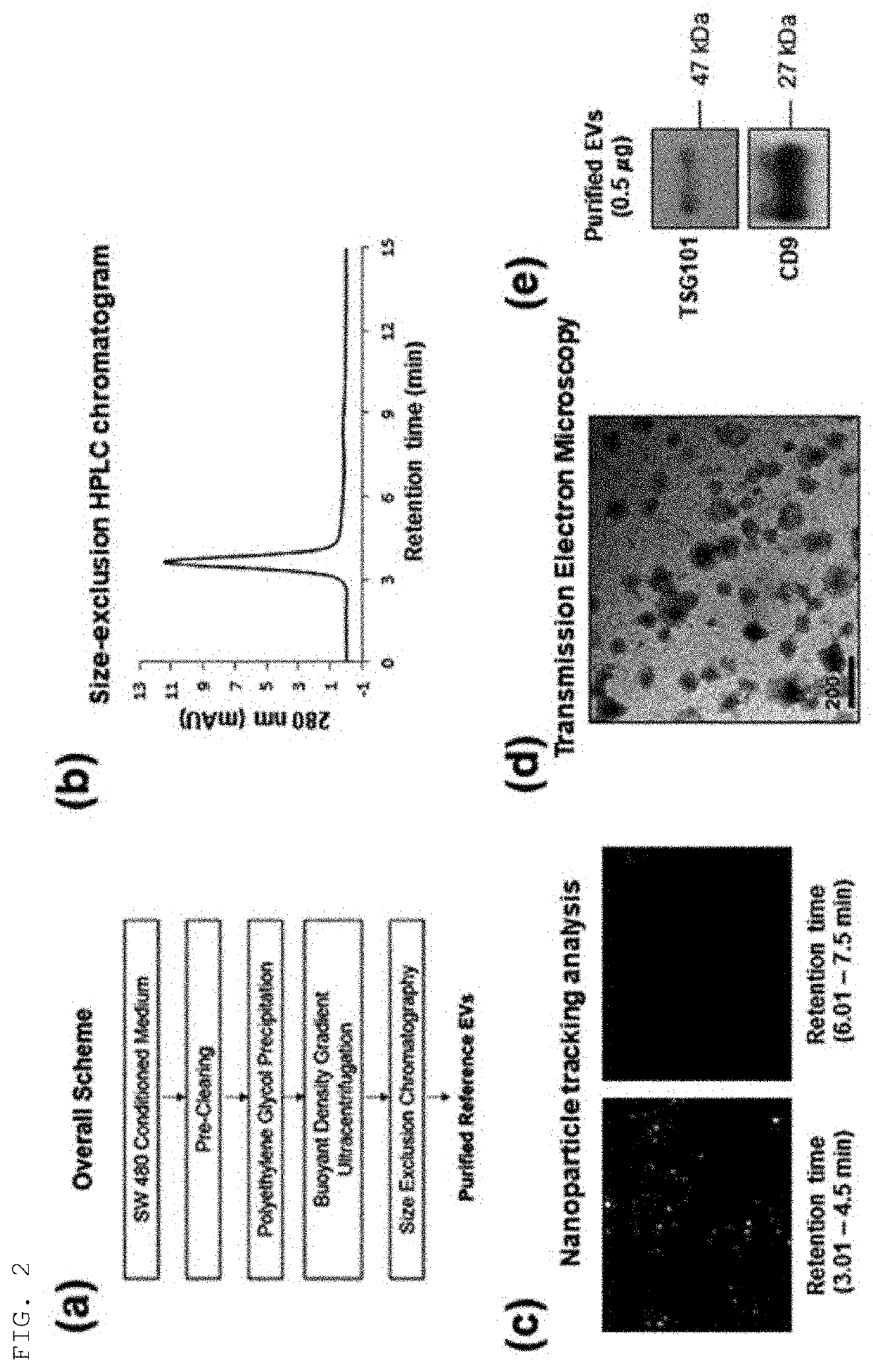
[0067]A colorectal cancer cell SW480 culture was centrifuged at 500×g for 10 min (repeated twice in total) to remove remaining cells and precipitates. The supernatant was again centrifuged at 2,000×g for 20 min (repeated twice in total) to remove precipitates.
[0068]To primarily purify and precipitate extracellular vesicles present in the supernatant, the supernatant was subjected to addition of an extracellular vesicle precipitation-inducing solution (8.4% polyethylene glycol 6000, 250 mM NaCl, 20 mM HEPES, pH 7.4), stored in a refrigerator for 16 hrs, and centrifuged at 12,000×g for 30 min to harvest the precipitated extracellular vesicles, which were then dissolved in HEPES-buffered saline (20 mM HEPES, 150 mM NaCl, pH 7.4).
[0069]To secondarily purify extracellular vesicles using density and buoyancy, the sample was mixed with Optiprep (to a final concentration of 30%), and placed at the lowest layer in an ultracentrifugati...
example 2
Isolation of Extracellular Vesicles Using Various Types of Cations with Several Concentrations
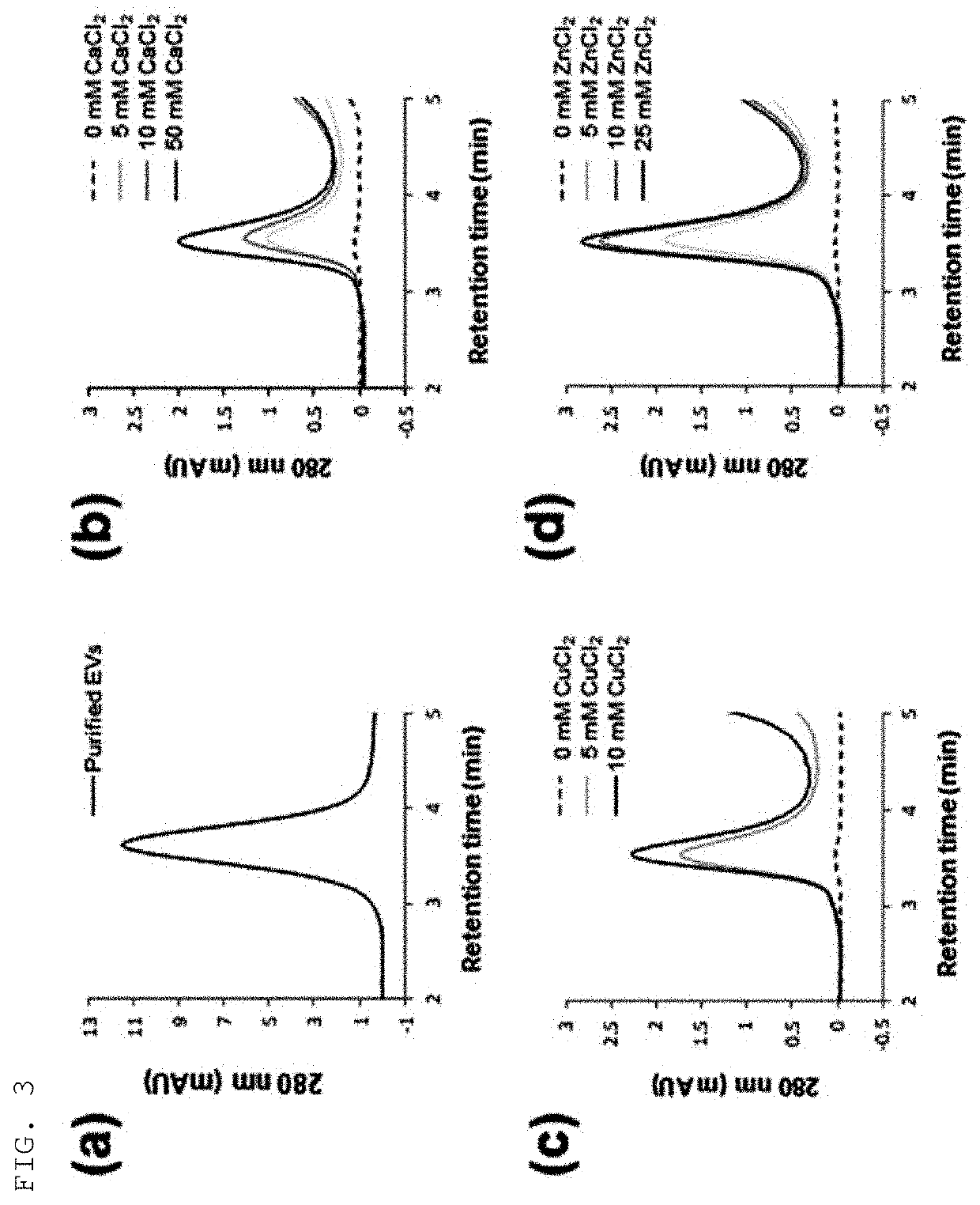
[0073]Colorectal cancer cell cultures were subjected to addition of various types of cations (Ca2+, Cu2+, Zn2+) with several concentrations, followed by mixing and then centrifugation at 3,000×g for 10 min, to thereby harvest precipitates, which were then dissolved in HEPES-buffered saline containing 50 mM EDTA. As a result of analysis of the isolated extracellular vesicles by size-exclusion chromatography using an HPLC system, the sample extracellular vesicles were detected at 3.6 min and are shown in FIG. 3A.
[0074]As for the treatment concentrations of calcium, copper, and zinc cations added to the colorectal cancer cell cultures, it was verified that the 280-nm absorbance band detected at 3.6 min increased in proportion with the concentration of cations, and these results are shown in FIGS. 3B to 3D. The absorbance bands in the respective types of cations and the absorbance band in the s...
example 3
Isolation of Extracellular Vesicles Using Copper Cations (Copper(II) Chloride)
[0075]Colorectal cancer cell cultures were subjected to addition of copper cations with several concentrations, followed by mixing and then centrifugation at 3,000×g for 10min, to thereby harvest precipitates, which were then dissolved in HEPES-buffered saline containing 50 mM EDTA. The extracellular vesicles isolated by the above method were investigated by nanoparticle tracking analysis and western blot analysis. For nanoparticle tracking analysis, the Nanosight LM10 instrument was used, and tracking and recording was made under the conditions of a camera level of 10 and a detection limit of 3 for 60 sec. For western blot analysis, the signal of CD9, which is a general extracellular vesicle marker, was analyzed after SDS electrophoresis.
[0076]As a result, it was shown in FIG. 4A that the yield of extracellular vesicles increased as the concentration of copper cations increased, and it was therefore confi...
PUM
| Property | Measurement | Unit |
|---|---|---|
| Concentration | aaaaa | aaaaa |
Abstract
Description
Claims
Application Information