Microspheres Containing Decellularized Donor Tissue and Their Use in Fabricating Polymeric Structures

a donor tissue and microsphere technology, applied in the field of microspheres containing decellularized donor tissue, can solve the problems of inability to re-generate functional cartilage akin to native tissue, high case dependence, and progressive degradation, and achieve the effect of enhancing migration, proliferation, and chondrogenesis of hmscs

Pending Publication Date: 2020-09-17
CHILDRENS HOSPITAL MEDICAL CENT CINCINNATI +1
View PDF1 Cites 3 Cited by
  • Summary
  • Abstract
  • Description
  • Claims
  • Application Information

AI Technical Summary

Benefits of technology

[0019]Disclosed herein is the controlled release of proteins, growth factors, cytokines, and proteoglycans from thermally stable matrix microsph

Problems solved by technology

Due to the limited proliferative capacity of chondrocytes and its avascular nature, injury to the osteochondral tissue can lead to progressive degradation—eventually to osteoarthritis and significant disability.
While current treatment options such as micro-fracture, autologous chondrocyte implantation and osteochondral autologous transfer improve the quality of life of some patients, they are not capable of regenerating functional cartilage akin to native tissue and are often highly case dependent.
Moreover, the end product is fibrocartilage that is mechanically inferior to the hyaline cartilage.
Currently there is no treatment for the restoration of large osteochondral injuries (OCI) that exceed the indications fo

Method used


Image

  • Microspheres Containing Decellularized Donor Tissue and Their Use in Fabricating Polymeric Structures
  • Microspheres Containing Decellularized Donor Tissue and Their Use in Fabricating Polymeric Structures
  • Microspheres Containing Decellularized Donor Tissue and Their Use in Fabricating Polymeric Structures

Examples


example 1

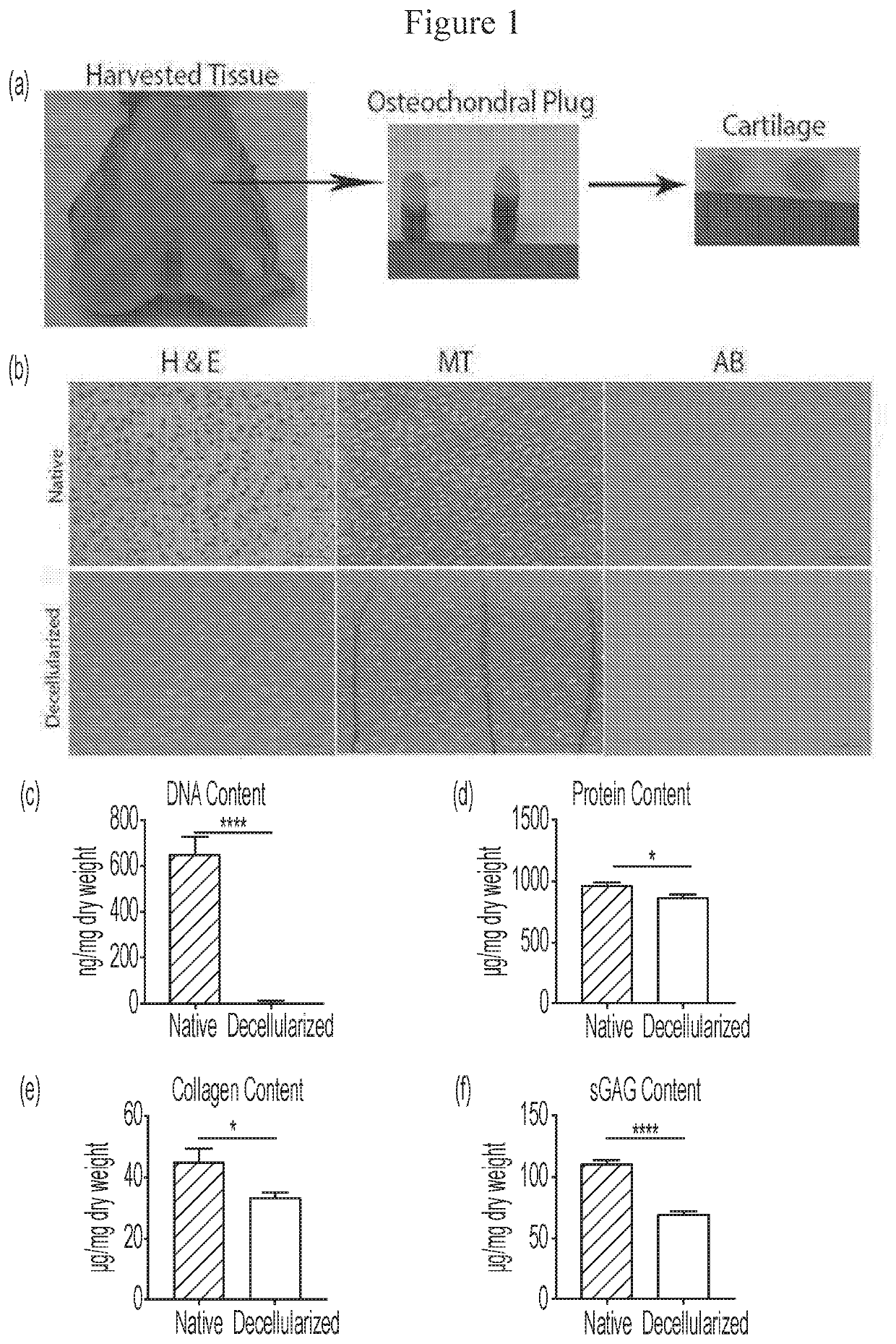
Harvest and Decellularization

[0110]The hind limbs of pigs were provided by the Center for Surgical Innovation, University of Cincinnati. The limbs were cleaned to remove excess blood and disinfected using aseptic techniques. The osteochondral plugs of 8 mm diameter were harvested from the femoral condyle using an Osteochondral Autograft Transfer (OAT) system.

[0111]The cartilage was then excised from the bone and were placed in sterile 15 mL conical tubes (Falcon) and stored at −80° C. until further use. The cartilage tissue was decellularized similar to the process previously described. See, Whitlock P W, et al., A naturally derived, cytocompatible, and architecturally optimized scaffold for tendon and ligament regeneration, Biomaterials 28: 4321-29 (2007).

[0112]Cartilage tissues were exposed to a series of solutions with continuous stirring in a rotating shaker (New Brunswick Scientific) at 200 rpm, 37° C. First, the tissues were immersed in 100 mL of nuclease free distilled water ...

example 2

c Analysis of Native and Decellularized Tissues

[0116]Fresh-frozen native and decellularized chondral tissues were placed in 10% phosphate-buffered formalin (Sigma) at room temperature for 24 h and were then processed for histology.

[0117]The tissues were dehydrated in a graded series of ethanol, soaked in xylene, embedded in paraffin and sliced with a microtome to obtain 5.0 μm thick, longitudinal sections. These sections were then mounted on slides and stained using hematoxylin and eosin (H&E), Masson's trichrome (MT) and alcian blue. The stained sections were then assessed under a light microscope (Nikon 90i).

example 3

al Evaluation of Native and Decellularized Tissues

[0118]The fresh-frozen native and decellularized cartilage tissues (n=10) were lyophilized (Labconco, Freeze Dry System, Kansas City, Mo.) for 72 h to achieve equivalent weight for DNA content evaluation. Total DNA was isolated from the cartilage tissues (Approximately 25 mg each) using a commercially available kit (DNeasy, Qiagen, Valencia, Calif.).

[0119]The total DNA content was measured by absorption at 260 nm using a spectrophotometer (Thermo Spectronic, Biomate 3, Rochester, N.Y.) and normalized to initial dry weight of the samples.

[0120]The lyophilized native and decellularized tissues were digested using pepsin (Sigma-Aldrich, St. Louis, Mo.)-HCl. In brief, 20 mg of the lyophilized samples was digested with 1 mg of pepsin in 0.01 N HCl (Sigma-Aldrich) for 48 h at room temperature under constant stirring. The partially digested samples were diluted with 10×PBS and 0.1 N NaOH.

[0121]Total protein content in the tissues was estima...


PUM

PropertyMeasurementUnit
Weight ratioaaaaaaaaaa
Structureaaaaaaaaaa

Abstract

Decellularized matrix microspheres comprising a polymeric material and a donor tissue are provided. Also disclosed are structures containing a plurality of decellularized matrix microspheres incorporating a first polymer and a donor tissue; and a second polymer, wherein the decellularized matrix microspheres and the second polymer are in the form of a filament. Methods of treating a tissue injury employing the matrix microspheres and structures described as well as their methods of manufacture are also provided.

Description

CROSS REFERENCE TO RELATED APPLICATIONS[0001]This application claims priority to U.S. Provisional Application No. 62 / 578,037 filed Oct. 27, 2017, the contents of which are incorporated herein by reference.FIELD[0002]The disclosure relates to microspheres containing decellularized donor tissue that remain functional after incorporation within a polymeric filament and their use in fabricating polymeric structures.BACKGROUND[0003]Articular cartilage is a highly complex connective tissue that covers the surface of the bones in synovial joints. Sophia Fox A J et al., The basic science of articular cartilage: structure, composition, and function, Sports Health 1: 461-8 (2009); and Temeno J S et al., Review: tissue engineering for regeneration of articular cartilage, Biomaterials 21: 431-40 (2000). Due to the limited proliferative capacity of chondrocytes and its avascular nature, injury to the osteochondral tissue can lead to progressive degradation—eventually to osteoarthritis and signif...

Claims


Application Information

Patent Timeline
IPC IPC(8): A61L27/18A61L27/36B33Y80/00B33Y70/10B29C64/118
CPCB29L2031/7532B33Y70/10A61L2430/06B29K2489/00B29K2467/046B29K2067/00A61L27/3612B33Y80/00B33Y10/00A61L27/18B29C64/118A61L27/3683B33Y40/10A61L27/38A61L27/54A61K38/18A61K35/32A61P19/08A61K35/28B33Y70/00A61L2300/622A61L2300/624C08L67/04
Inventor LIN, CHIA-YING JAMESGRUBER, STACEYWHITLOCK, PATRICK W.GHOSH, PAULOMI
Owner CHILDRENS HOSPITAL MEDICAL CENT CINCINNATI
Why Eureka
Try Eureka