
Enzymatic method for removal of cryoprotectants from cryopreserved animal cells

a technology of cryoprotectant and cryopreserved animal cells, which is applied in the field of enzymatic method for removal of cryoprotectants from cryopreserved animal cells, can solve the problems of destroying biological cells and tissues, unable to discover blood-borne diseases with a latency period longer than 14 days, and unable to detect blood-borne diseases in donors, etc., to achieve controllable conversion rate

Inactive Publication Date: 2001-04-17
COOK GEN BIOTECH
View PDF7 Cites 1 Cited by
  • Summary
  • Abstract
  • Description
  • Claims
  • Application Information

AI Technical Summary

Benefits of technology

The present invention relates to a method of removing chemical compounds which protect cells (cryoprotectants) during low temperature preservation (cryopreservation). Typically such cryoprotectants are small molecular weight molecules such as glycerol, propylene glycol, ethylene glycol or dimethyl sulfoxide (DMSO). More particularly, this invention is directed to the use of specific enzymes to convert the cryoprotectant compounds into forms which cannot move across the cell membrane and enter the cell (are plasma membrane impermeable). Therefore, when applied to the cells, the enzymatic activity will shift the chemical potential of the cryoprotectants inside and outside the cells, causing the cryoprotectants to flow out of the cell, in turn become enzymatically modified, and thereby removed from the cells. The rate of conversion can be controlled.
This approach differs fundamentally from all previous methods in that it utilizes enzymes to effect cryoprotectant removal thereby minimize osmotic damage due to cell swelling and thus enhances the viability of cryopreserved cell populations.

Problems solved by technology

Moreover, diseases carried in blood with a latency period longer than 14 days may not be discovered in the donor until the blood has been placed into a patient.
The survivability of viable cells and tissues using prior art freezing methods is often quite low.
Freezing conditions are relatively harsh and thermal shock or other phenomena such as ice crystal formation often destroy biological cells and tissues.
Ericsson reports that his method increases the fertility of a sperm sample by enhancing the environmental (the ratio of total sperm to motile sperm) and the viability (progressiveness of motility of the motile sperm) factors affecting the fertility of a sample, but his method does not improve the population (motile sperm count) factor which is possibly the most critical.
Most of these methods are, however, complex, cumbersome and expensive to utilize.
A disadvantage of the aforementioned methods resides in that low temperature preservation of the cells and tissues is accomplished by the ice crystallization process.
As ice forms in the solution surrounding the cells or tissues, electrolytes and other solutes become progressively concentrated, quickly reaching concentrations which are damaging to the cells.
However, the cryoprotectants themselves can cause osmotic damage to the cell during their addition and removal.
The cryoprotectant removal process and associated cell swelling is particularly damaging.

Method used


Image

  • Enzymatic method for removal of cryoprotectants from cryopreserved animal cells
  • Enzymatic method for removal of cryoprotectants from cryopreserved animal cells
  • Enzymatic method for removal of cryoprotectants from cryopreserved animal cells

Examples


example 1

Use of Glycerol Kinase to Remove Glycerol from Cells in a Single Step Process

Glycerol kinase (GK) catalyzes the following reaction: ##EQU2##

Glycerol kinase is found widely in nature (Thorner and Paulus, 1973). In microorganisms GK is involved in the utilization of glycerol as a carbon source. In mammals, the enzyme in involved in fat and sugar metabolism (Kida et al., 1973). GK phosphorylates glycerol exclusively to L-[[alpha]]-glycerophosphate. The reaction is essentially irreversible and Mg.sup.2+ is required (Zwaig and Lin, 1966, Hayashi and Lin, 1967; Thorner and Paulus, 1973). The activity of this enzyme is characterized in terms of the ability of the enzyme to convert 1.0 .mu.M glycerol to L-glycerol, 3-phosphate / min at 25.degree. C.

Many cells, such as red blood cell, used widely in transfusion medicine, are cryopreserved using 1-3 M concentrations of glycerol. One of the major rate limiting factors using current protocols for freezing red blood cells, is the time it takes to ...

example 1a

Determination of Human RBC Cell Volume in the Presence of Glycerol Phosphate

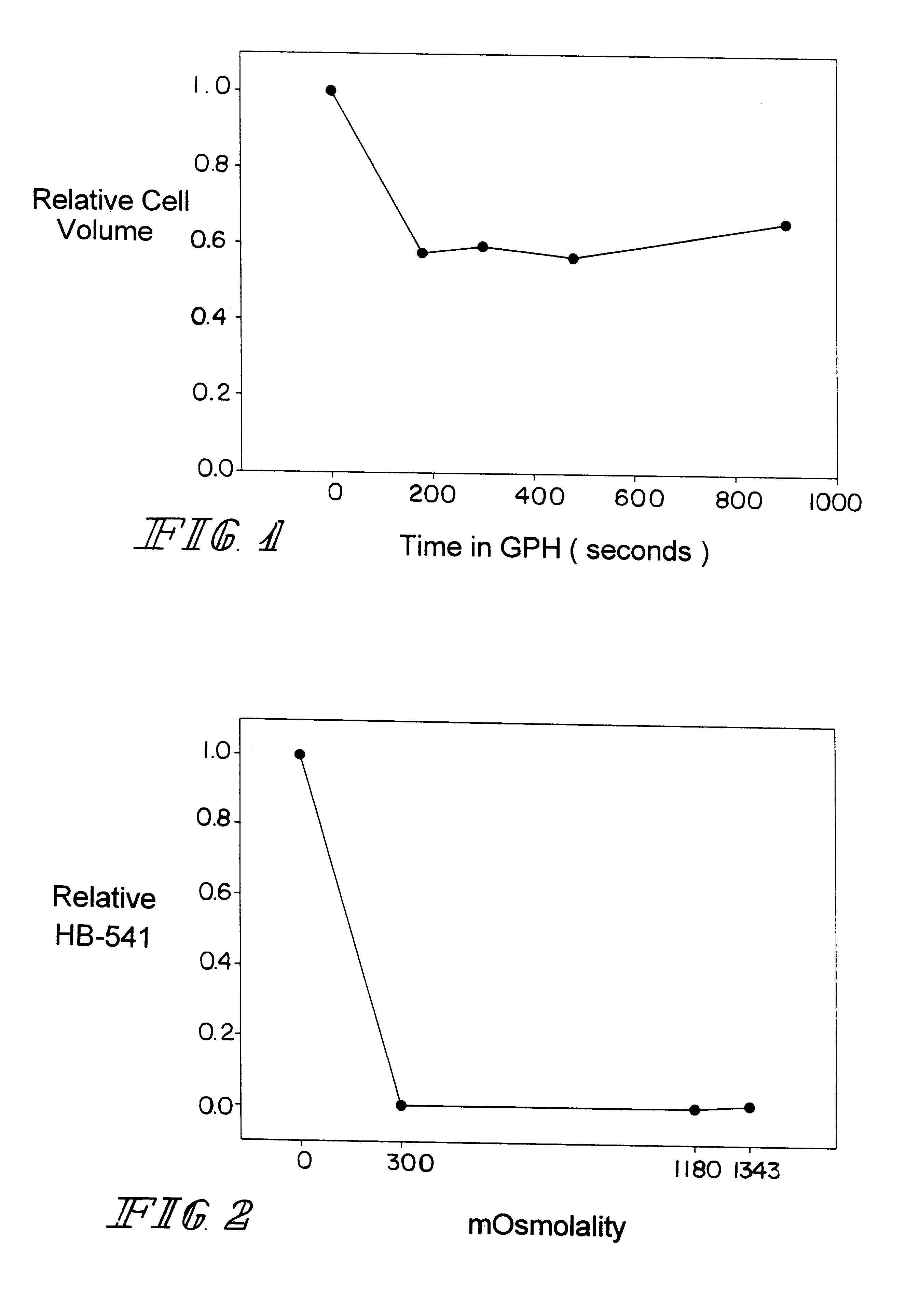
Media: 950 mg of L.alpha.GPH were placed into 6.42 ml of isotonic phosphate buffered saline (PBS) to yield a 0.4 M concentration of L.alpha.GPH. The corresponding osmolality was 1340.+-.3 mOsm.

Experimental procedure: Human red cells were abruptly exposed to the 0.4 M L.alpha.GPH solution and the cell volume over time was determined using a modified Coulter counter apparatus. The results are shown in FIG. 1. Human RBC volume was reduced to 58% of their isosmotic volume within 3 minutes and the cell volume remained reduced for 15 minutes. These data demonstrate that L.alpha.GPH is impermeable to the human red cell plasma membrane.

example 1b

Determination of Human RBC Lysis in the Presence of L.alpha.GPH

Media: Reagent grade distilled water (negative control; 100% lysis expected), isotonic PBS (positive control, 0% lysis expected) and 1180 and 1340 mOsm glycerol-phosphate solutions were used.

Experimental procedure: Human red cells were exposed for 20 minutes to one of the four treatment media listed above. After 20 minutes, the cells were placed into a spectrophotometer and absorbance at 541 nm measured to determined the amount of hemoglobin in free solution as a measure of cell lysis. The distilled water treatment was used as the negative control and assumed to produce 100% cell lysis. The other values were normalized to this value. The results are shown in FIG. 2. No cell lysis was observed in either the isosmotic PBS control (positive control) or the L.alpha.GPH solutions. These data indicated that concentrations of L.alpha.GPH to remove glycerol from human red cells are not damaging.


PUM

PropertyMeasurementUnit
Temperatureaaaaaaaaaa
Concentrationaaaaaaaaaa
Permeabilityaaaaaaaaaa

Abstract

A method to remove cryoprotectants from cryopreserved biological cells and tissues is described. Enzymes are used to convert compounds used as cryoprotectants for cells during cryopreservation to membrane impermeable products. This process effectively removes the cryoprotectant chemicals from the cells prior to their use in transfusion, transplantation, insemination or other applications, without causing osmotic damage due to cell swelling. Methods are described for subsequent removal of the enzymes and enzyme conversion products from the cell and tissue preparations.

Description

The present invention relates to a method of removing chemical compounds which protect cells (cryoprotectants) during low temperature preservation (cryopreservation). More particularly, this invention is directed to the use of specific enzymes to convert cryoprotectant compounds into products which are not transported across the cell membrane. The enzymes convert cryoprotectants in solution and thereby work to shift the chemical potential of the cryoprotectant resulting in osmotic flow of cryoprotectants out of the cells and into the enzyme solution.BACKGROUND OF INVENTIONFresh biological cells such as sperm, red blood cells, platelets and the like, are typically viable for but a short period of time in vitro. Nevertheless, it is commercially and medically significant that such cells are available for use long after they have been collected from donors, sometimes several months or even years later. Various cryopreservation methods, have been developed to preserve biological cells fo...

Claims


Application Information

Patent Timeline
IPC IPC(8): A01N1/02
CPCA01N1/0221A01N1/0205
Inventor CRITSER, JOHN K.WOODS, ERIK J.
Owner COOK GEN BIOTECH
Learn More