

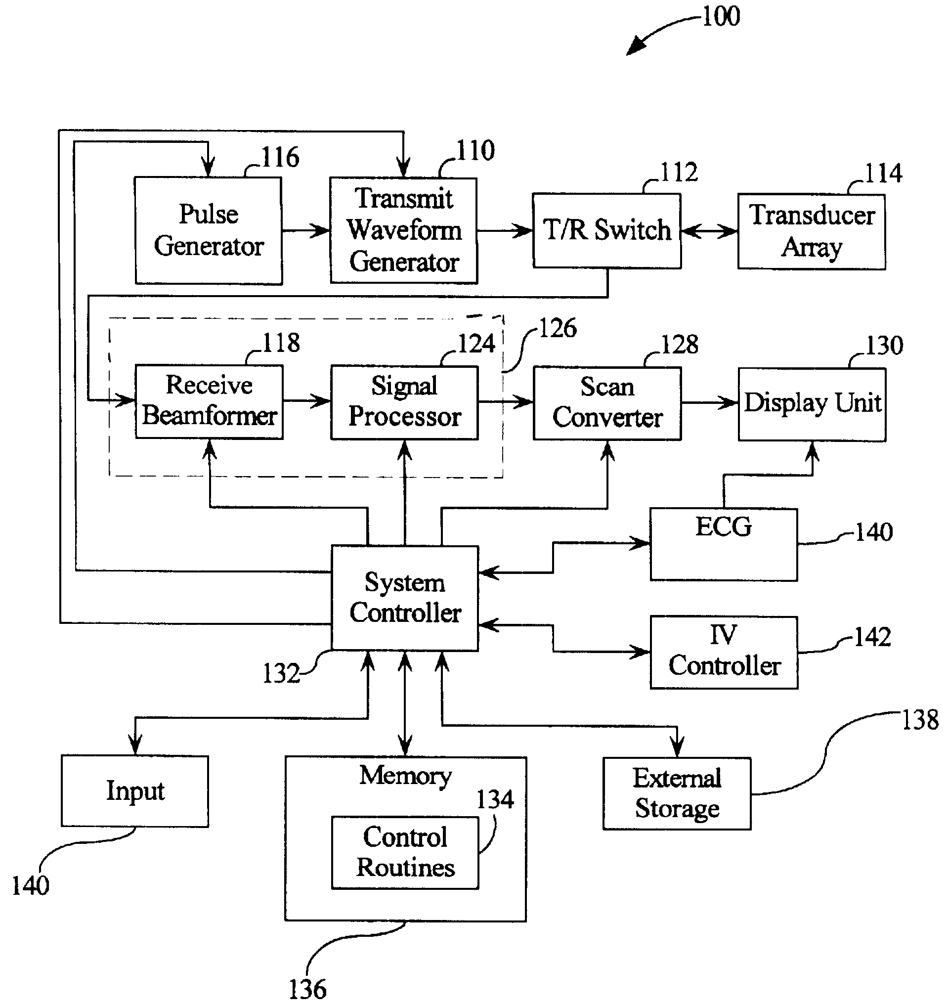
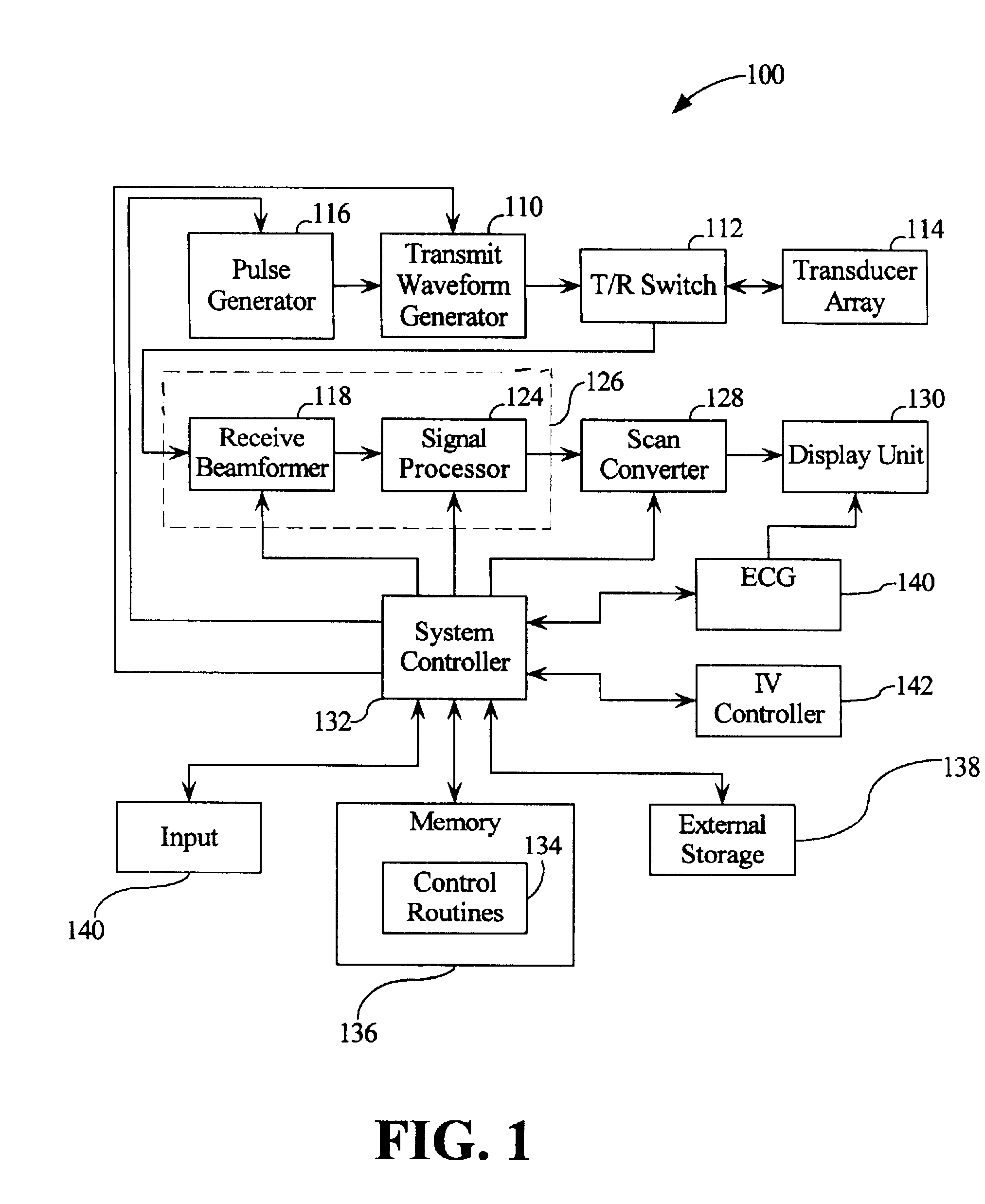
Automated ultrasound system for performing imaging studies utilizing ultrasound contrast agents
an automated, imaging study technology, applied in tomography, reradiation, instruments, etc., can solve the problems of coronary artery obstruction, still quite a challenge, complex parametric changes, etc., to achieve rapid change of imaging modes and parametric information, imaging studies utilizing contrast agents, and the effect of reducing the difficulty of patients
- Summary
- Abstract
- Description
- Claims
- Application Information
AI Technical Summary
Benefits of technology
Problems solved by technology
Method used
Image

Examples
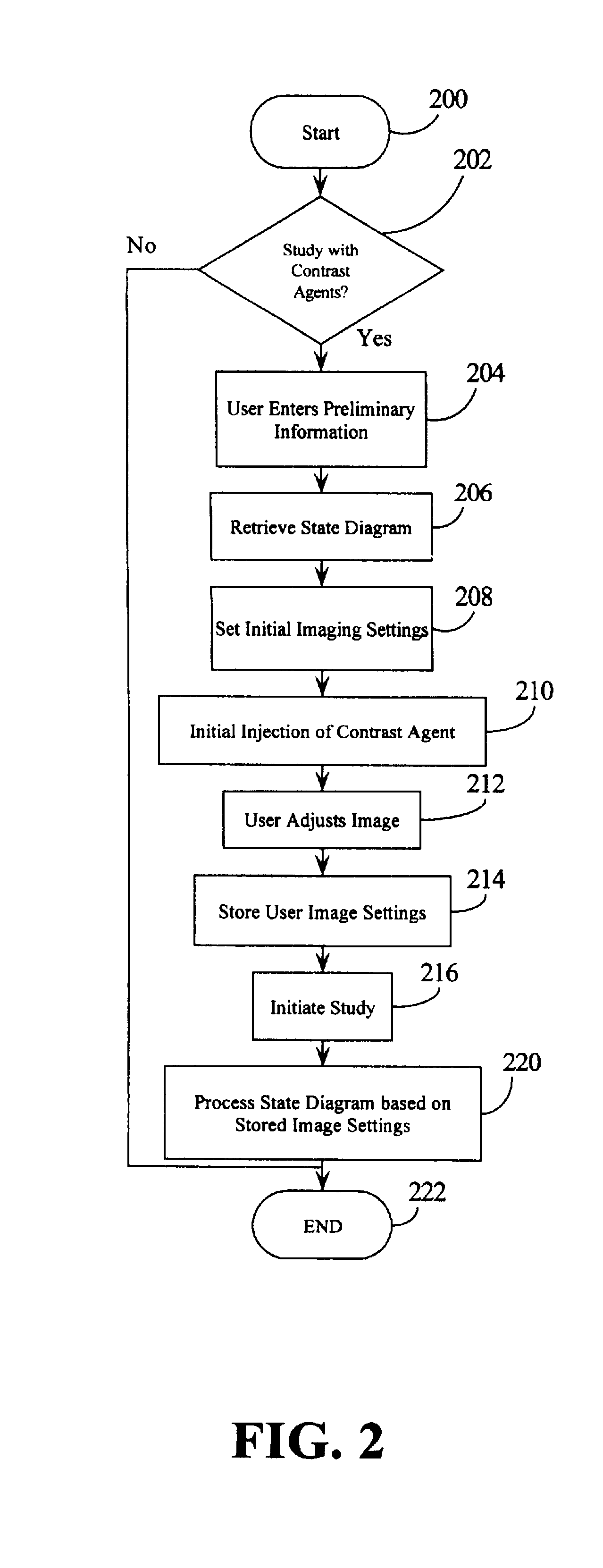
example 2
Contrast Study for Left Ventricular Opacification / Myocardial Perfusion
1. User determines need for contrast based on prior examination and also performs baseline imaging to determine appropriate windows for MCE and appropriate windows for LVO. The user then starts the routine in step 200 and confirms the use of contrast agent in step 202.
2. User inputs information before beginning the study including the following:
Type of MCE study (e.g., Dobutamine, exercise, Dipyridamole, Adenosine infusion, Adenosine bolus, etc.)
Agent to be used (e.g., Optison, Definity, Levovist, etc.)
Method of Administration (e.g., infusion or bolus)
Patient information (weight, height, gender, body type, level of difficulty in each imaging plane, etc.)
imaging views will MCE be done on, which imaging planes will LVO be done on.
Desired Output for quantification (e.g., myocardial blood volume, flow-volume curve, coronary flow reserve, etc.).
3. Based on this information, the system selects a state diagram, in step 2...
PUM
 Abstract
Description
Claims
Application Information