This process, however, is very
time consuming and quite subjective, and frequently results in poorly shaded restorations.
These cameras are designed to be versatile and able to collect measurements in tight places often found in the mouth; however, they do not preserve the color fidelity—that is, they do not collect the true color—of the object measured.
Unfortunately, conventional intraoral cameras suffer two problems: distance sensitivity due to illumination geometry and
color discrimination error due to sensor limitations.
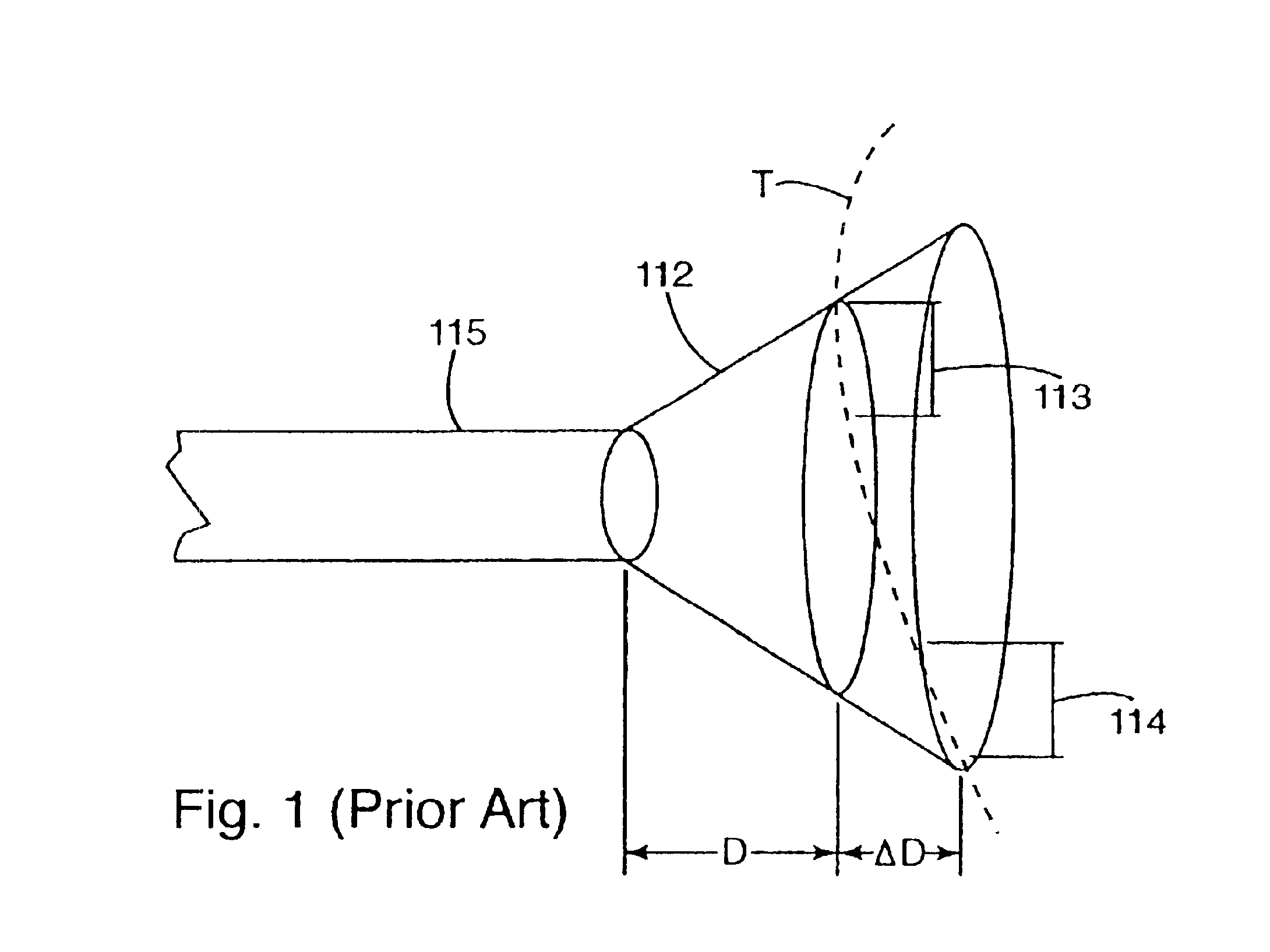
Although
fiber-optic illumination is useful for providing high levels of illumination and is compatible with small measuring probe tips, a drawback of any small illumination source that illuminates a larger area is that the projected beam must be divergent light.
It is known that when the distance change to the illumination source is significant with respect to the distance to the source, the illumination output varies significantly, creating what is called non-uniform illumination.
Particularly with objects positioned close to the
fiber optics, certain regions of the object are non-uniformly illuminated because the light from the illumination source rapidly diffuses as it travels away from the source.
Moreover, when multiple sources of light are used to illuminate an object, the object may be non-uniformly illuminated in different regions.
Given this non-uniform illumination, a
color sensor, sensing the light reflected from the tooth, will invariably collect inconsistent color information from region to region.
With non-uniform illumination, conventional intraoral cameras critically rely on illumination source positioning which can not be maintained in practical use.
This results in significant errors affecting tooth shade determination.
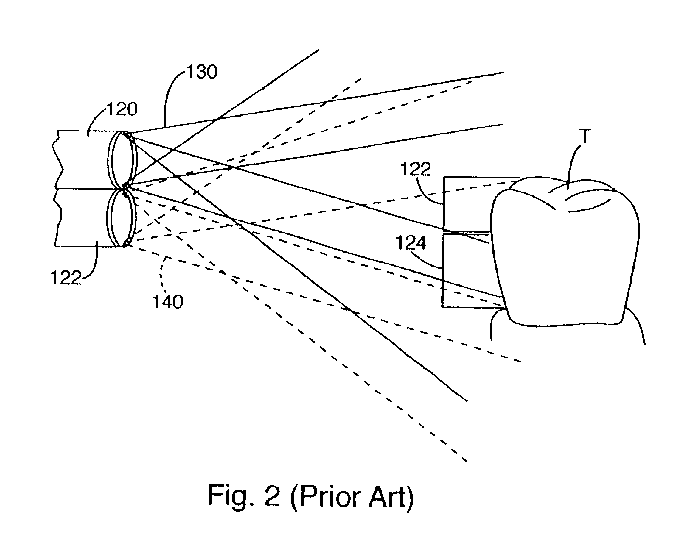
A limitation of this method of illumination is that the illuminant intensity is maximized at the intersection of the two projected beams.
Often, significant portions of the measured area are not illuminated by both beams and hence have a lower and unpredictable illumination value.
Given this non-uniform illumination, a
color sensor, sensing the light reflected from the tooth, will invariably collect inconsistent color value information front region to region.
In addition to non-uniform illumination, today's intraoral cameras utilize
color filter array (CFA) image sensors that frequently contribute to inaccurate
color measurement because the filter array is applied to the image.
Although RGB sensors offer a means to collect
color data for a tooth, that data often is not an accurate representation of the true color or distribution of color on the tooth.
CFAs do not accurately measure color primarily because of two factors: pixel spacing separation and poor color fidelity.
Moreover,
prosthesis manufactured from this measurement data collected with an RGB sensor will not accurately reflect the true color of each point on the tooth.
These design goals come at the cost of color fidelity.
More specifically, today's RGB CFA collects selected wavelengths of light impinging on them, but they also incidentally collect unwanted wavelengths in the process.
This lack of rejection of light outside of the wavelengths of interest degrades color fidelity to an unacceptable level for accurate
color measurement.
Due to
signal detection problems caused by pixel spacing and poor color fidelity, CFA-type sensors are not accurate enough for satisfactory determination of tooth shade.
Although these protective sheaths prevent spread of contaminants, their functionality is limited exclusively to this sanitary purpose.
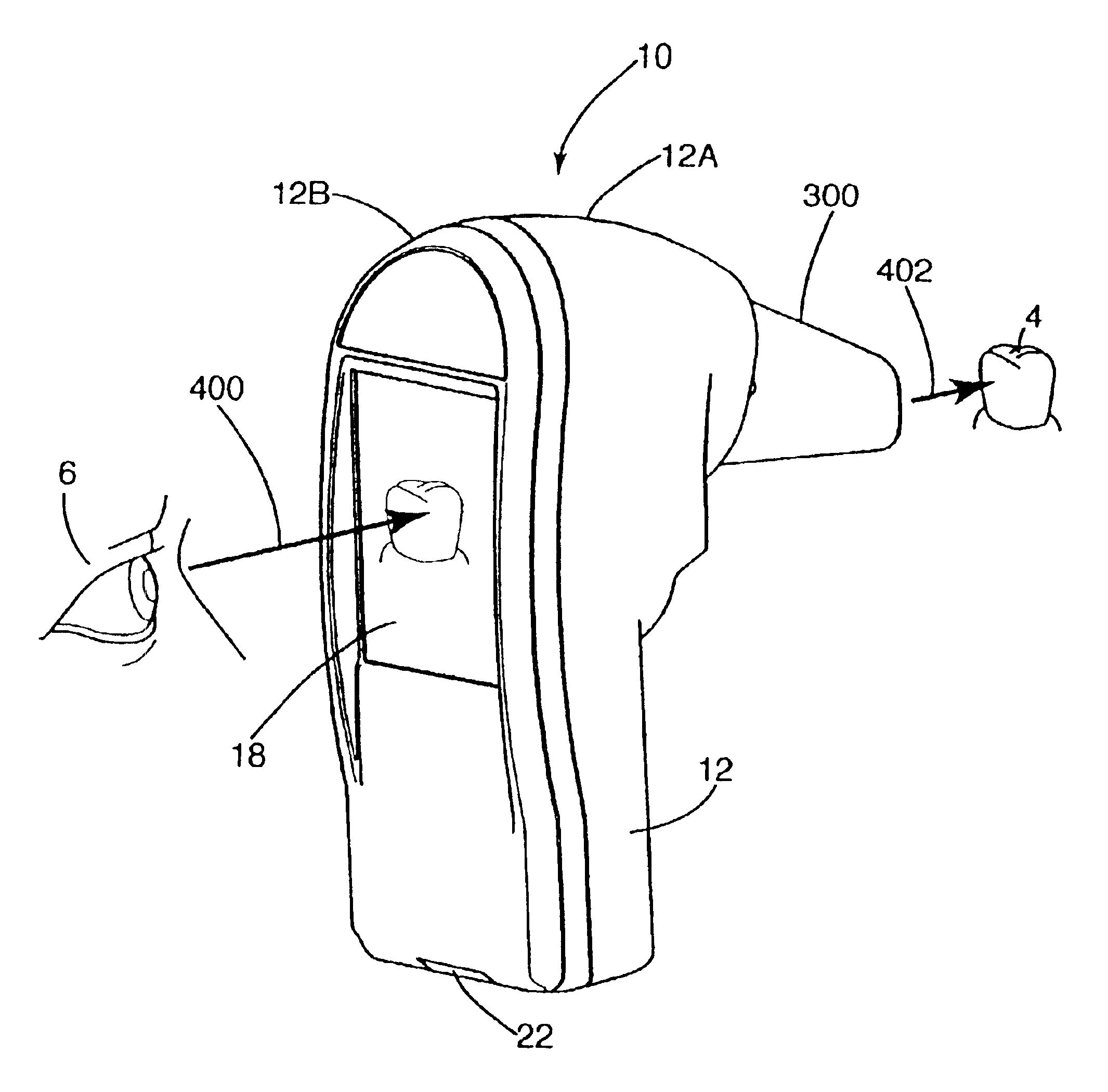
The drawback in collecting images of a tooth with these conventional probes is that the dentist must look back and forth from the probe to the monitor to insure the probe is positioned over the tooth to obtain the desired image on the monitor.
This, of course can cause unneeded
frustration in aligning the probe to collect measurements of the tooth.
In many instances, intraoral cameras or parts thereof intentionally or accidentally come into contact with a patient's intraoral cavity thereby transmitting contaminants including infectious agents,
saliva and / or food debris to the device.
This is often a tedious task, as the cameras include a plurality of buttons that are difficult to clean around and / or
fiber optic bundles that are nearly impossible to sterilize without damaging the optical characteristics of the fibers because sterilization agents enter the fiber
optics and degrade illumination or sensing capabilities.
Accordingly, prior art camera users must
exercise time-consuming care in operating and cleaning these cameras.
This process is very costly if multiple prosthetic replacements must be produced to create a satisfactory match.
Moreover, this process consumes the time of patients who may come in for repeated visits before a matching
prosthesis is created.

