Methods and compositions to prevent formation of adhesions in biological tissues

a biological tissue and adhesion technology, applied in the field of methods and compositions to prevent adhesion formation in biological tissues, can solve the problems of material not remaining in intimate contact with the treated biological surface for a sufficient period of time, infertility, pain, etc., and achieve the effects of preventing postoperative adhesion, reducing the extent of tumor cell metastasis, and protecting injured blood vessels

Inactive Publication Date: 2006-04-18
CALIFORNIA INST OF TECH
View PDF14 Cites 50 Cited by
  • Summary
  • Abstract
  • Description
  • Claims
  • Application Information

AI Technical Summary

Benefits of technology

[0017]The compositions can be applied to isolated tissue or to tissue during surgery or by means of a catheter or other less invasive device, for example during angioplasty or atherosclerosis surgery. The compositions are useful for blocking adhesion and immune recognition and thereby aid in the prevention of postoperative adhesions, protection of injured blood vessels from thrombosis and intimal thickening relating to restenosis, and decrease the extent of metastasis of tumor cells. The materials can be used as barriers to prevent the interaction of one cell or tissue with another cell or tissue, and as carriers for bioactive species. A wide variety of biological and nonbiological surfaces, with different geometries, can also be coated with these polymeric materials.

Problems solved by technology

These adhesions can have severe consequences including pain, bowel obstruction, and infertility.
Adhesions are scar tissue bridges that often connect to opposing organs, for example, restricting organ movement and function as well as causing pain.
In some cases, the materials did not remain in intimate contact with the treated biological surface for a sufficient period of time.
The most popular of these materials are oxidized regenerated cellulose and expanded polytetrafluoroethylene, but use of these barriers has resulted in questionable efficacy, due to the requirement for extensive surgical manipulation, sometimes including additional surgery, nonadherence of the barrier to the surface, and immune response.
These materials may work by preventing tissue damage in the first place and / or by “floating” the tissues, but the materials can make surgical manipulation difficult and all of the materials have shown questionable efficacy.

Method used


Image

  • Methods and compositions to prevent formation of adhesions in biological tissues

Examples


example 1

The Reductive Amination of Lysine with Phenylboronic Acid

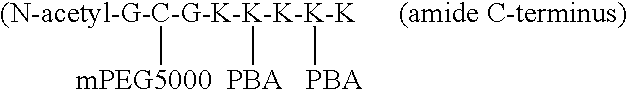
[0063]N-acetyl-Gly)-Cys(Trt)-Gly-(Lys(Mtt)5 was synthesized on 2.7 g of PEG-polystyrene resin with 0.2 mmol / g functionality (the resin used yields an amide C-terminus). The lysine protecting groups were deprotected by soaking the resin in 1% trifluoroacetic acid, 5% triisopropylsilane in DCM for about 3 minutes then filtering. This was repeated ten times and the material was then washed with DCM. The lysine was then desalted by adding 10 times excess triethylamine in DCM and then rinsing with DCM. The peptide-resin, 1.05 equivalents of 4-formylphenylboronic acid and 10 equivalents of sodium triacetoxyborohydride (based on the number of lysines present) were placed under argon and dissolved with anhydrous DCM so the peptide was 2 mM (not all of the reactants dissolved completely). The reaction was kept under argon and stirred for 17.5 h. Then the solution was rinsed with DCM, rinsed copiously with water saturated with sodium bi...

example 2

Grafting of PEG to the Peptide-PBA Compound

[0064]2.67 g of thiopropyl Sepharose™ 6B gel was swollen in water and then rinsed copiously with water. A 0.1 M phosphate, 0.5 M NaCl, 5 mM EDTA, 1 mM TCEP, pH 7.00 buffer was sonicated. 10.7 mL of buffer and 0.46 g of TCEP were added to the gel, placed under argon, and swirled for 1 hour. The result was rinsed copiously with the previously mentioned buffer, poured into a chromatography column, and greater than 3 bed volumes of buffer was flowed through by gravity. The column was then capped. The peptide-PBA compound of Example 1 was added to 0.09 g of TCEP, and 6 mL of buffer. The solution was stirred and 300 μL was reserved (as “free mL of buffer 3 times. All eluents were combined and dialyzed against phosphate buffered saline 2 times, saline 7 times, and water 4 times, for a total of 5 days. The dialyzed solution was filtered through a 0.2 μm Acrodisc™ PF syringe filter and lyophilized.

[0065]The results of NMR and Ellman's assays demonst...


PUM

PropertyMeasurementUnit
solubilityaaaaaaaaaa
adhesionaaaaaaaaaa
tissue adhesionaaaaaaaaaa

Abstract

The invention discloses materials that adsorb readily to the surfaces of body tissues in situ and provide a steric barrier between such tissues, so that tissue adhesions, which typically form following surgical procedures, are minimized. These materials contain a polymer of hydrophilic molecules such as polyethylene glycol (PEG) bound to a polymer that spontaneously adsorbs to biological tissue such as phenylboronic acid (PBA). The PEG-PBA co-polymer can be formed in a variety of geometries. The materials can also be used to coat prosthetics and other implants.

Description

CROSS REFERENCE TO RELATED APPLICATION[0001]This application is a divisional of U.S. Ser. No. 09 / 140,781 filed on Aug. 27, 1998, now U.S. Pat. No. 6,596,267 by Jeffrey A. Hubbell, Natalie D. Winblade, and Donald L. Elbert and entitled “Methods and Compositions to Prevent Formation of Adhesions in Biological Tissues”, which claims priority to provisional patent application Ser. No. 60 / 056,854 filed on Aug. 27, 1997 by Jeffrey A. Hubbell, Natalie D. Winblade, and Donald L. Elbert and entitled “Steric Protection of Biological Surfaces by the Adsorption of Copolymers Containing Poly(ethylene glycol) and Phenylboronic Acid”.STATEMENT REGARDING FEDERALLY FUNDED RESEARCH[0002]The U.S. Government has certain right in this invention by virtue of a National Science Foundation Graduate Fellowship to Natalie Winblade and NIH Grant number HL56297 to Jeffrey Hubbell.FIELD OF THE INVENTION[0003]The present invention relates to methods and polymeric compositions for preventing the formation of tiss...

Claims


Application Information

Patent Timeline
Patent Type & Authority Patents(United States)
IPC IPC(8): A61F13/00A61K47/30A61P17/02A61P29/00A61P35/00A61K47/00A61K31/77A61L31/00A61K31/78A61K31/79A61K31/80A61L27/34A61L29/00A61L31/06A61L31/10C08L101/02
CPCA61K31/77A61K31/79A61K31/80A61L27/34A61L31/06A61L31/10A61K31/78C08L71/02C08L101/02Y10S977/931Y10S525/937A61P17/02A61P29/00A61P35/00
Inventor HUBBELL, JEFFREY A.WINBLADE, NATALIE D.ELBERT, DONALD L.
Owner CALIFORNIA INST OF TECH
Why Eureka
Try Eureka