Phototherapy device and method of use

a technology of phototherapy and edema, which is applied in the field of phototherapy devices and methods of use, can solve the problems of no possibility of affecting the physiological processes of tissue cells, no cure effect for the whole class of diseases accompanied by metabolic disorders, and no cure effect for the whole class of diseases

Inactive Publication Date: 2006-07-25
HART BARRY M +1
View PDF7 Cites 200 Cited by
  • Summary
  • Abstract
  • Description
  • Claims
  • Application Information

AI Technical Summary

Problems solved by technology

When the body has been injured either by accident or through medical procedures there is going to be a period of time where inflammation and edema will set into the effected tissue area.
From this hyper-activity, the capillary and postcapillary networks become flooded and expand causing hyperemia.
They, however, provide no possibility to affect the physiological processes in tissue cells, or their effects are mediated by a number of uncontrolled factors.
Thus, it cannot produce a curing effect for the whole class of diseases accompanied by metabolic disorders.
However, the stimulation used for the purpose is thermal.
This method, however, is applied only to stop bleeding and to increase blood coagulability through use of wavelengths ranging from 5.0 to 1.1 mu.m that are not effective for curing the whole set of medical indications typical to the entire class of diseases in question.
Besides, the produced effects provide no increase to the curing efficiency compared to the optimum curing effect for diseases caused by disorders of metabolic, regenerative and enzymatic processes in tissues.
However, the efficiency of this method is rather low, since the irradiation is performed through the skin site located directly over the affected region of mucosa and is unable to produce an optimum effect on metabolic, enzymatic, and regenerative processes in tissues.
The given method of treatment provides a curing effect after a large number of irradiation sessions, however, complications are observed in the form of tissue necrosis and edema that decrease the efficiency of treatment by lowering the level of effects on tissues' regenerative, enzymatic, and metabolic processes.
This is caused by the fact that the shallow penetration of the emission is unable to activate all the potentials of tissue structures across their entire thickness.
Besides, activation and optimization of processes in tissues is also not equally effective for different types of tissues, different locations of affected tissues (deep or shallow), and different types of diseases.
This inc

Method used


Image

  • Phototherapy device and method of use
  • Phototherapy device and method of use
  • Phototherapy device and method of use

Examples


Embodiment Construction

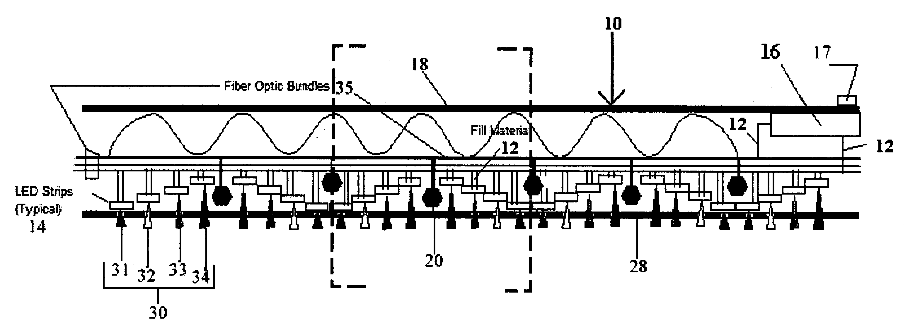
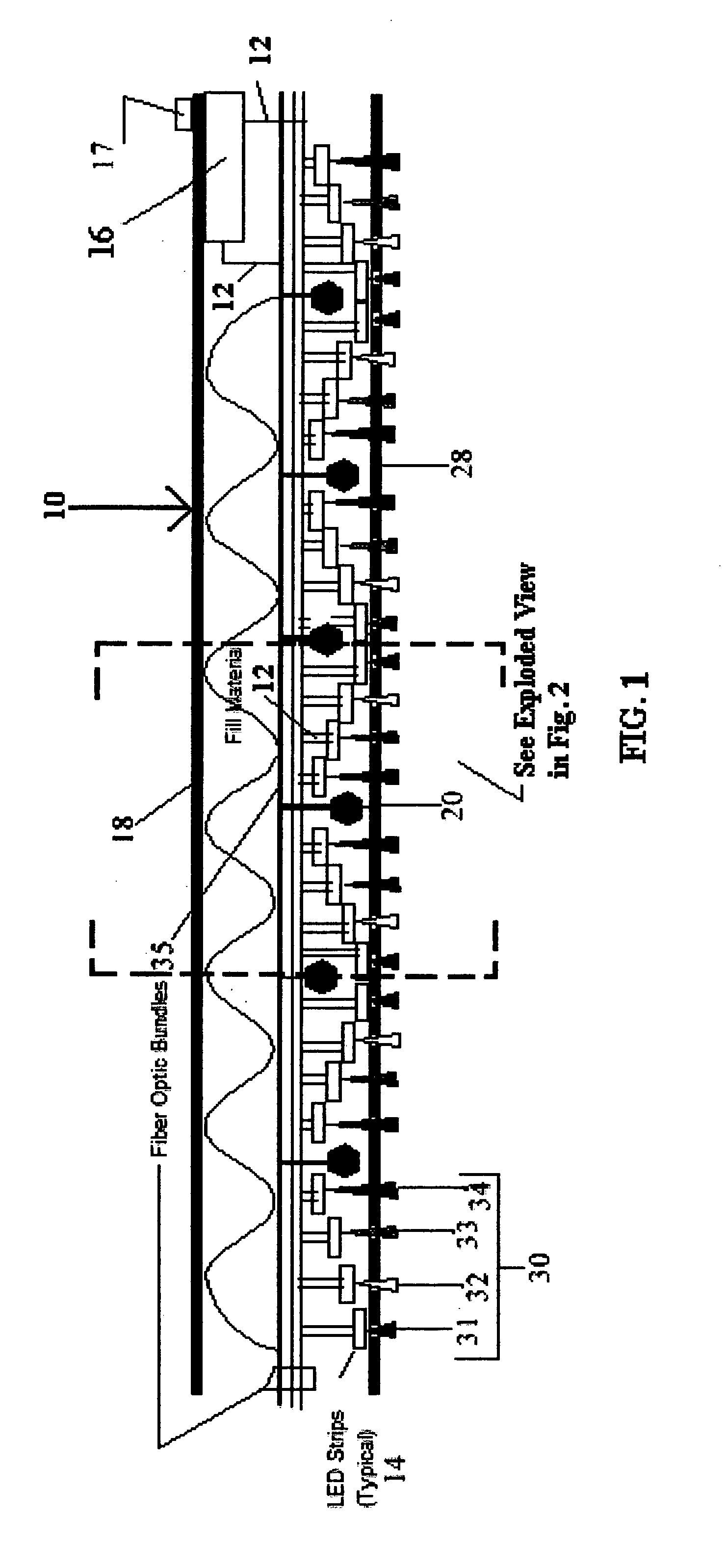
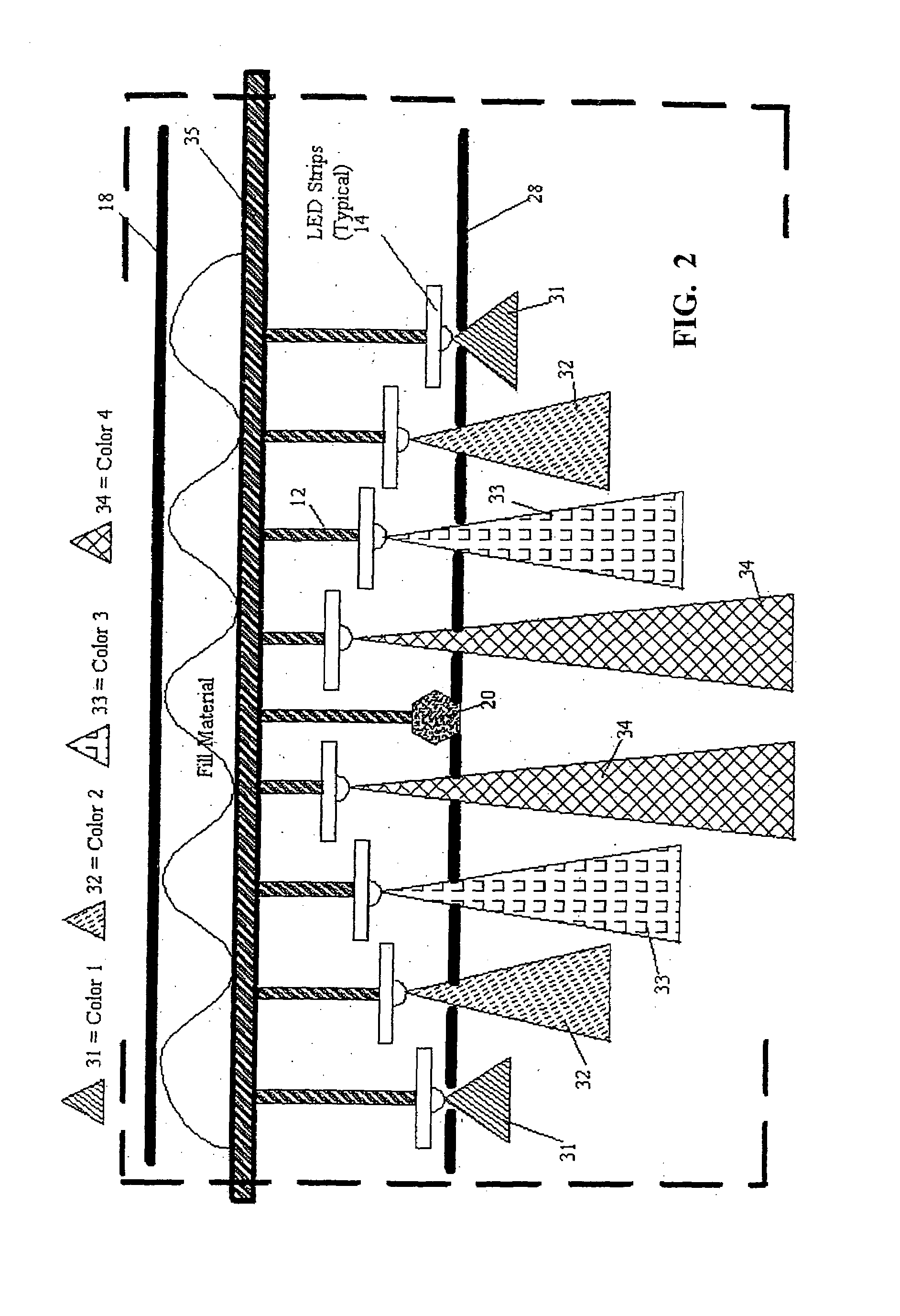
[0047]The Joint / Tissue Inflammation Therapy and Monitoring Device or “JIT-Mon” is made of a neoprene elastic material like that found in wetsuits. Sizing of the device will be made with Velcro type straps allowing for an easy and comfortable fit. Each JIT-Mon has strategically located Light Emitting Diodes (LED's) with calibrating wavelengths and modulated light frequencies to allow for controlled heat / energy and muscular therapy to an area of inflammation. Additional monitoring devices, using optical fiber, photodetector and photoresin technologies, are integrated into the JIT-Mon device and are supported with customized software and hardware. This information and technology allows a physician or therapist to monitor and record vital information such as blood flow in the area, skin temperature and moisture to an external-monitoring device.

[0048]By surrounding the injured and inflamed areas with an elastic fitted device, which applies controlled heat / energy using Light Emitting Diod...


PUM


Abstract

A device is provided, in direct skin contact, surrounding an injured area for the treatment, reduction of joint inflammation, edema and excitation of neural and muscular stimulation associated with human and mammal tissues. This therapeutic light source includes a multiplicity of light emitting diodes (LED's) found in the ranges of 350 nm to 1000+ nm and fiber optic connections. A neoprene type material or other non-allergenic material is used to set the LED's and fiber optics in layers consisting of contact with the skin to few centimeters from the skin tissue. Distance will vary from contact or near contact with devices to several millimeters of separation. Each LED array is independently controlled allowing for optimal modulation of light frequencies and wavelengths. Technology is integrated allowing for biomedical feedback of tissue temperature and other statistical information. A low voltage, portable power supply, is integrated into the device as well as an analog/digital, input/output connection device. The design will be created for continuous wear, flexibility and comfort.

Description

RELATED APPLICATIONS[0001]This application is claims benefit of Provisional Application No. 60 / 361,161 filed Mar. 4, 2002, which is herein incorporated by reference.FIELD OF THE INVENTION[0002]Design and composition of a device or devices to be used in a method for the stimulation of human and animal tissue, allowing for the reduction of inflammation and edema to joints, tissue and nerve bundles associated with trauma. This device consists of the implementation of light-emitting diode technologies such as modulated light frequencies and wavelengths but not limited to this technology. In addition, the design incorporates custom software programming and engineering allowing for diagnostic interpretation, biomedical recording and patient statistical / historical medical events in “real time” mode. This will allow for the data to be transmitted via telemetry or “direct connect” to other diagnostic equipment.PRIOR ART REFERENCES[0003]U.S. Patent DocumentsPatent #DateAuthor4932934June 1990D...

Claims


Application Information

Patent Timeline
IPC IPC(8): A61N5/06
CPCA61N5/0616A61N2005/063A61N2005/0663A61N2005/0647A61N2005/0652A61N2005/0645
Inventor HART, BARRY M.MALAK, HENRYK
Owner HART BARRY M
Why Eureka
Try Eureka