Method for electro-permeabilization of individual cellular and organellar structures and use thereof

Inactive Publication Date: 2006-09-19
CELLECTRICON
View PDF22 Cites 59 Cited by
  • Summary
  • Abstract
  • Description
  • Claims
  • Application Information

AI Technical Summary

Benefits of technology

[0013]Furthermore, the present invention distinguishes itself from prior art in that electroporation with single-cell or subcellular spatial resolution is accomplished by applying the electric field through nanometer- and micrometer-diameter electrodes with extremely short interelectrode distances. The electrodes are controlled individually by high-graduation micromanipulators, allowing precise focusing of the electric field between the electrodes. Electroporation of individual cells and individual organelles can thereby be accomplished. Electroporation can with the present invention be performed in such a way that only a target cell is permeabilised and not its adjacent neighbour. Also, individual cellular processes can be electroporated. Even spatially well-defined intracellular domains with a targeted class of organelles can be held under a localised electric field with this invention, thereby enabling transfer of polar solutes organelles. Applications of electroporation of organelles include alterations of the mitochondrial genome. It is well known that mutations in the mitochondrial genome can lead to a multitude of diseases, and that gene therapy can potentially be of major importance. So far, however, mitochondria has to be isolated from the cells before transfer of the new genefragment into the mitochondria can be performed. Then, the mitochondria has to be reinserted into the cell. The technique according to the invention makes it possible to directly insert genes into the mitochondria when they are contained inside a cell. This is a significant advancement over traditional schemes for transfection of mitochondria.
[0014]In

Problems solved by technology

One of the major disadvantages with the known techniques is that they are not ap

Method used


Image

  • Method for electro-permeabilization of individual cellular and organellar structures and use thereof
  • Method for electro-permeabilization of individual cellular and organellar structures and use thereof
  • Method for electro-permeabilization of individual cellular and organellar structures and use thereof

Examples


example 1

Electroporation of Progenitor Cells from Mature Rat Brain to Incorporate a Fluorescent Marker

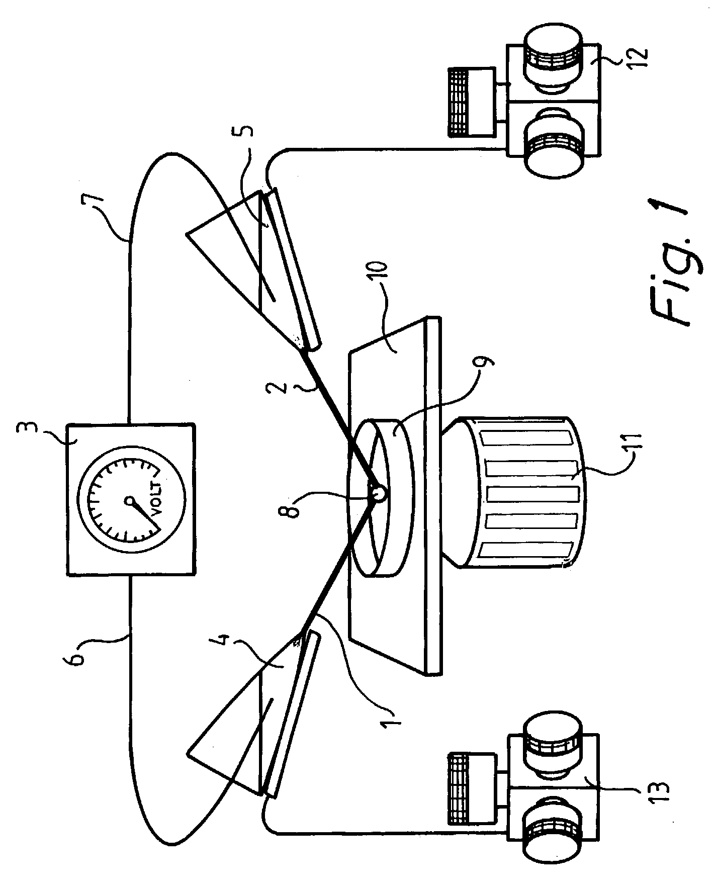
[0070]Progenitor cells were cultured according to standard procedures (T. D. Palmer, J. Ray, F. H. Gage, Mol. Cell. Neurosci., 6, 474–486 (1995)) and plated onto number 1, 1-inch circular coverslips coated with poly-D-ornithine and lamilin. The cells were allowed to stand overnight to adhere to the glass surface. The cells were incubated in a humid atmosphere at 37° C. with 5% CO2 and 95% air. For the electroporation, the cell dishes were mounted into a circular low-rim polycarbonate holder using mounting grease. The polycarbonate holder with the cover slip was mounted on the stage of an inverted microscope (Leica DM IRB). The cells were held in a HEPES-buffered saline containing 137 mM NaCl, 0.4 mM MgCl2, 0.64 mM KH2PO4, 3.0 mM NaHCO3, 0.41 mM MgSO4, 5.4 mM KCl, 20 mM HEPES, 1.26 mM CaCl2, and 5.5 mM D-glucose (pH adjusted to 7.4 with NaOH). This HEPES buffer was supplemented with fluoresce...

example 2

Electroporation of Intracellular Organelles in Progenitor Cells from Adult Rat Brain Using Extracellular Electrodes

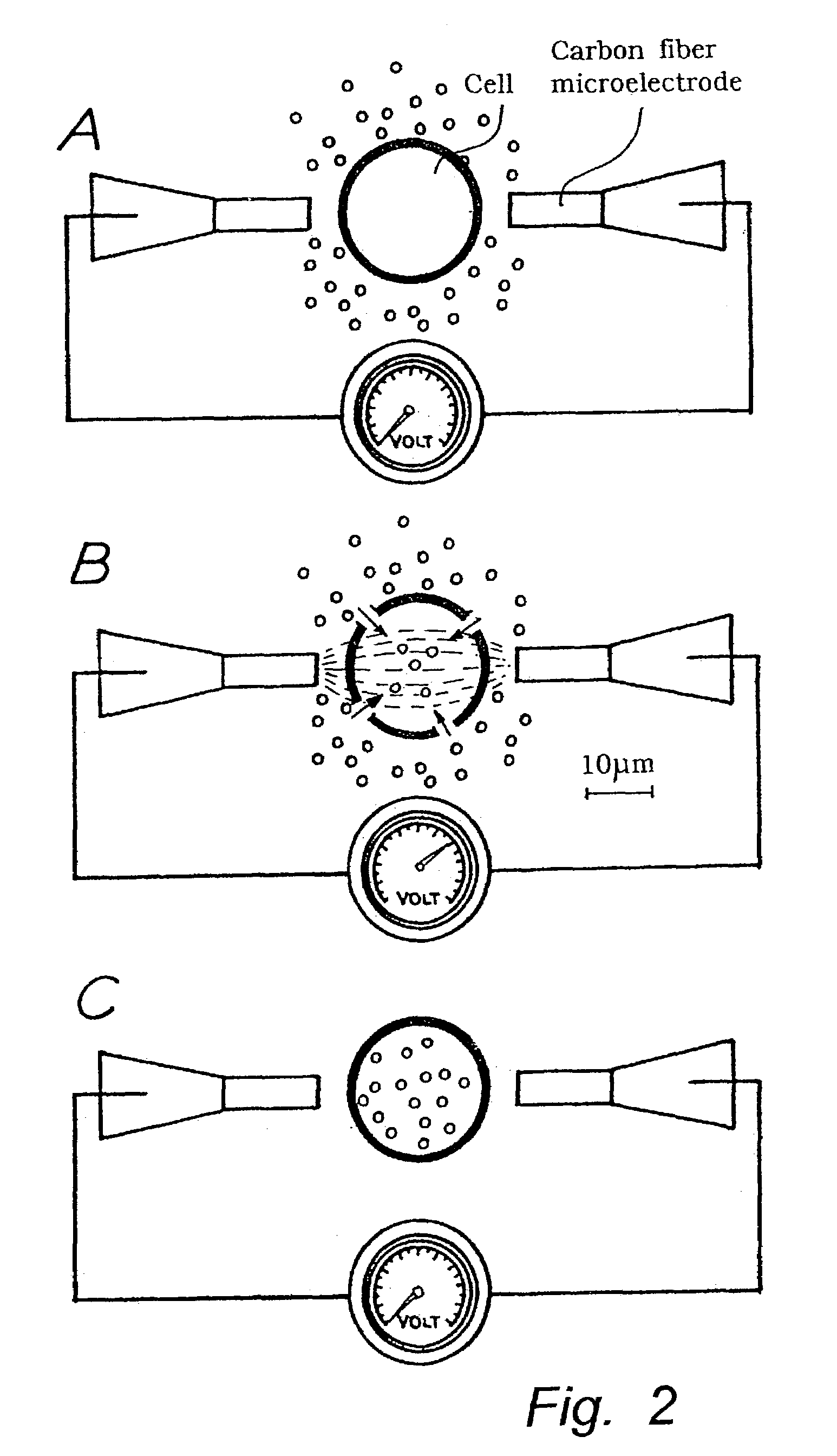
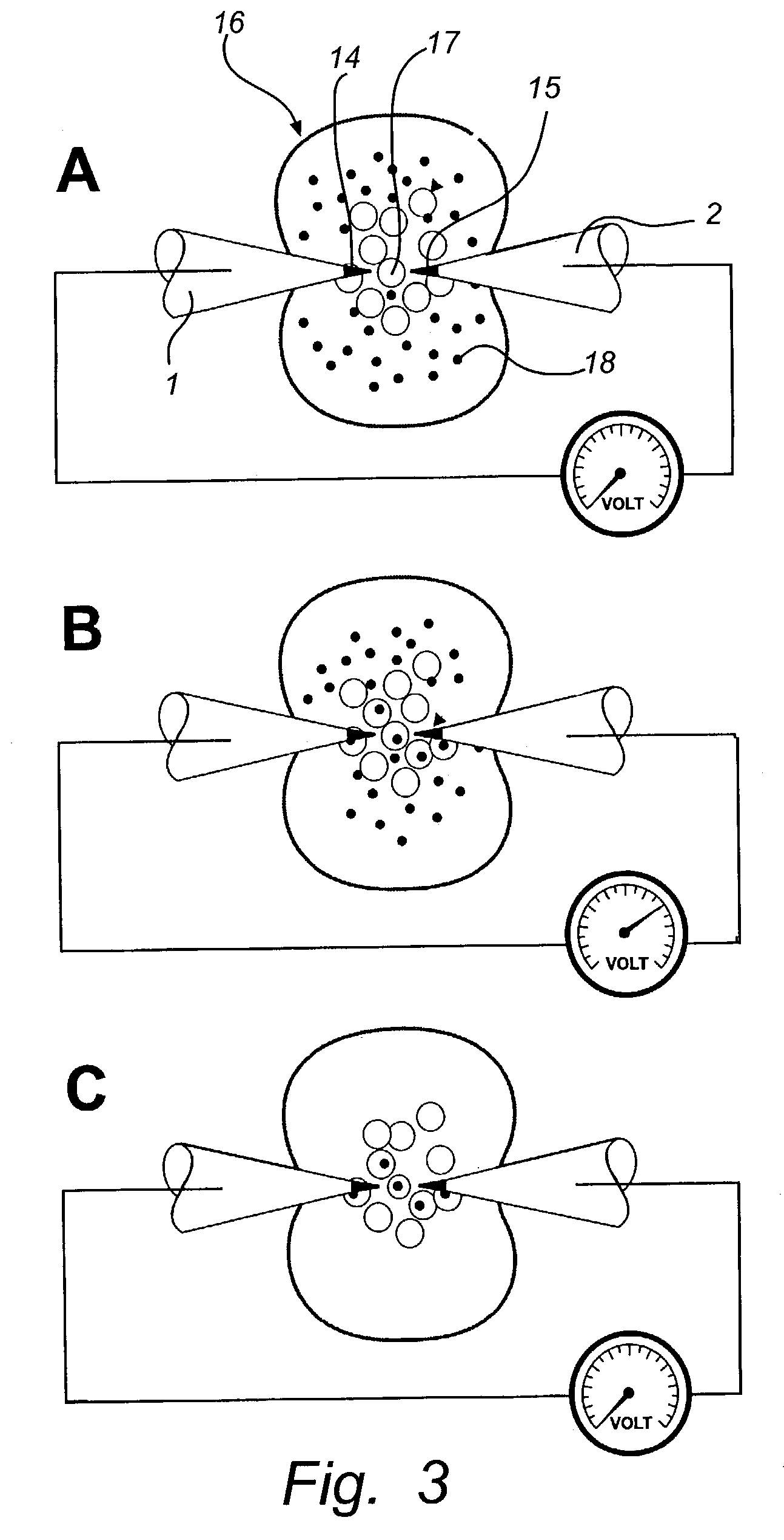
[0073]The experimental set-up from example 1 was repeated for in situ electroporation of organelles in single progenitor cells. Specifically, fluorescein was introduced into single cells by ten 0.5-Hz superthreshold field applications resulting in potential shifts at the cell membrane of 1.6 V±0.07 V (range 1.5–2.4 V), and compared to cells electroporated at low membrane potentials of 0.5–1.0 V. Following electroporation, the extracellular dye-containing media was replaced by a Hank Hepes solution. The result of electroporation at high voltages is shown in FIG. 7A, where punctuate fluorescence is observed in the exo-nuclear cytoplasmic region, but not in the nuclear region which contains a smaller number of organelles. In total, eighteen out of twenty cells electroporated in this protocol displayed a similar staining pattern, reminiscent of what is observed using an org...


PUM

PropertyMeasurementUnit
Lengthaaaaaaaaaa
Lengthaaaaaaaaaa
Diameteraaaaaaaaaa

Abstract

The invention relates to a method for permeabilization of a cell structure consisting of a single cell, an intracellular structure or an organelle comprising the following steps: (a) microelectrodes, preferably two carbon fiber electrodes or hollow fiber electrodes, are provided, (b) the microelectrodes are connected to a power supply, (c) the electrodes, individually controlled by high-graduation micromanipulators, are placed close to the cell structure at an appropriate inter-electrode distance, and (d) a highly focused electric field of a strength sufficient to obtain electroporation is applied between the electrodes. The method may be used in order to transfer cell impermeant solutes, such as drugs or genes, into the cell structure or out of the cell structure, in biosensors, in the treatment of tumours and neurodegenerative diseases and in the study of biophysical processes.

Description

RELATED APPLICATIONS[0001]This application is a continuation of U.S. Ser. No. 09 / 557,979, filed Apr. 25, 2000 now U.S. Pat. No. 6,521,430 which claims priority to PCT / SE98 / 02012, filed Nov. 6, 1998 in English which claims priority so SE 9704076-0, filed Nov. 6, 1997. The entirety of these applications is incorporated by reference herein.FIELD OF THE INVENTION[0002]The present invention relates to a highly spatially resolved method for permeabilisation of cell structures, organelle structures and cell-like structures in order to transfer compounds into or out of the structures. It also relates to use of this method.BACKGROUND OF THE INVENTION[0003]During the last two decades there has been a tremendous growth in experimental methods that allow for biochemical and biophysical investigations of single cells. Such methods include patch clamp recordings which can be used for measurement of transmembrane currents through a single ion channel (O. P. Hamill, A. Marty, E. Neher, B. Sakman, F...

Claims


Application Information

Patent Timeline
IPC IPC(8): C12M1/42C12M1/00A61N1/30A61N1/32C12N13/00
CPCC12N13/00C12M35/02A61N1/327A61N1/306
Inventor ORWAR, OWELUNDQVIST, ANDERSERIKSSON, PETERCHIU, DANIEL
Owner CELLECTRICON
Why Eureka
Try Eureka