
Imaging of drug accumulation as a guide to antitumor therapy

a technology of drug accumulation and imaging, applied in the field of radiolabeled antitumor drugs, can solve the problems of time required and toxicity of particular antitumor drugs

Inactive Publication Date: 2006-11-28
HEALTH & HUMAN SERVICES THE GOVERNMENT OF THE US REPRESENTED BY THE SEC
View PDF3 Cites 4 Cited by
  • Summary
  • Abstract
  • Description
  • Claims
  • Application Information

AI Technical Summary

Benefits of technology

[0039]Non-invasive, external imaging methods to visualize the location of a drug in the body avoid the need for biopsies and also have the capability of scanning large areas of the body, indeed, the entire body if necessary.
[0040]Since a drug is only effective if it reaches the desired site of action, a method to determine the location of the drug after administration allows monitoring of the potential effectiveness of drug administration. Antitumor drugs in particular are often toxic, and monitoring could reduce the time required to find the most effective and targeted therapy. Administration of a radio-labeled antitumor drug (preferably, the chemotherapeutic drug itself) which has been radio-labeled, preferably with a positron emitter, can provide external monitoring of drug accumulation in the tumor and / or normal host tissue by use of imaging technologies, preferably with a PET scanner.
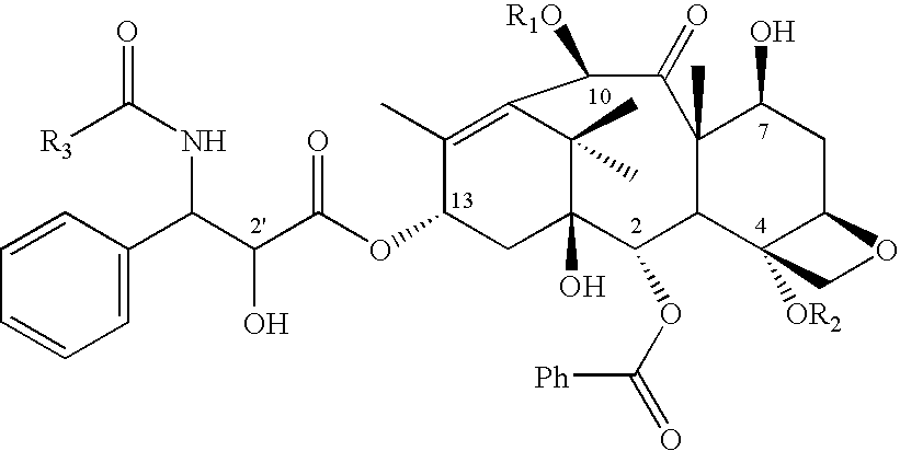
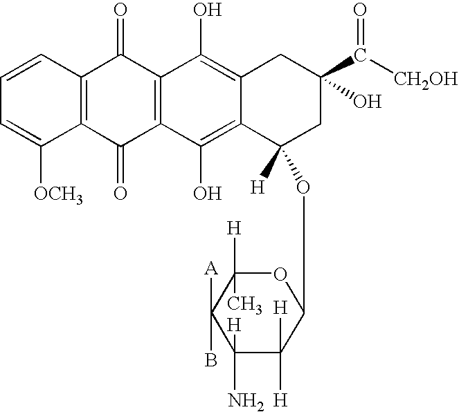
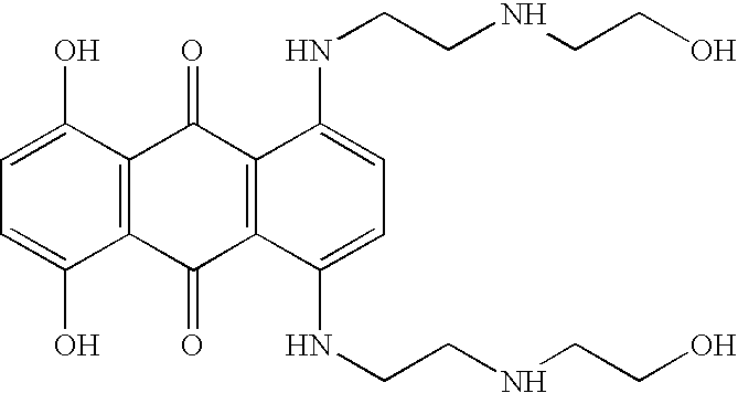
[0046]One goal of the present invention is to avoid exposing patients to toxic drugs that have no potential for benefit. The screening procedure of the present invention allows a rapid determination of whether a given tumor will be likely to respond to a particular drug. Such a rapid determination is important for other reasons, including the tendency of tumors to become refractory with time. Thus, immediate imaging with 11C-paclitaxel, 11C-docetaxel, 11C-doxorubicin, 11C-topotecan, 11C-mitoxantrone or other radio-labeled antitumor drugs has the advantage of selecting patients with tumors that are likely to respond to therapy with the particular drug that is used. Further, the impact of various doses and schedules for delivery can be monitored in situ at the actual tumor under treatment conditions. This is important since levels of MDR and other modifiers of drug efficacy are often induced or altered during the course of therapy. It is within the scope of the present invention to re-test patients as often as therapeutically or diagnostically necessary to assess the changing efficacy of treatment.

Problems solved by technology

Antitumor drugs in particular are often toxic, and monitoring could reduce the time required to find the most effective and targeted therapy.

Method used


Image

  • Imaging of drug accumulation as a guide to antitumor therapy
  • Imaging of drug accumulation as a guide to antitumor therapy
  • Imaging of drug accumulation as a guide to antitumor therapy

Examples


example 1

Synthesis of 11C-Paclitaxel

[0099]10-Deacetylpaclitaxel (6 mg) (commercially available) in pyridine (0.5 ml) was reacted with chlorotriethylsilane (0.1 ml) for 1 hour at 60° C. to yield 7,2′-di-(triethylsilyl)-10-deacetylpaclitaxel. This molecule is the immediate precursor for radiolabeling; it is stable at room temperature and can be stored until needed.

[0100]To a solution of (50 μg, 60 nmol) of 7,2′-di-(triethylsilyl)-10-deacetylpaclitaxel, was added dimethylaminopyridine solution (90 μl of 300 mg / ml in methylene chloride), and of tert-butyl diphenyl chlorosilane (20 μL). Acetyl chloride (10 μl of a 1:5000 solution in methylene chloride, 25 nmol) was then added and the mixture heated to 105° C. for 10 minutes. The silyl groups were removed within 2 minutes by adding of methylene chloride (400 μl) and tetrabutylammonium fluoride solution (50 μl of 1 M solution in tetrahydrofuran). Verification of the identify of the product via HPLC and mass spectrometry was obtained by comparison w...

example 2

Alternative Synthesis of 11C-Paclitaxel

[0103]Benzoyl chloride (8 μl of 1:1000 dilution in acetonitrile) is added to a solution of paclitaxel primary amine (50 μg) (commercially-available) in acetonitrile (200 μl). After 2 minutes at room temperature, 60% recovery of product was obtained.

[0104]Conversion of this procedure to radio-synthesis requires preparation of 11C-benzoyl chloride, which is known in the art by following typical Gringard reaction schemes. Mathews W B, Burns H D, Danals R F, Rabert H T, Naylor E M. Carbon-11 labeling of a potent, nonpeptide, AT1-selective angiotensin-II receptor antagonist, MK-996. J. Labelled Compounds and Radiopharmaceuticals 1995; XXXVI:729–37. For 11C-paclitaxel, purification may be accomplished by HPLC or by adding acidic water and methylene chloride.

example 3

Synthesis of 11C-Docetaxel

A. Preparation of Docetaxel Primary Amine

[0105]The starting material for the preparation of 11C-docetaxel is docetaxel primary amine, which is not available commercially. The free amine is prepared by removal of the tert-butyl carbonyl group from docetaxel using formic acid or other methods known in the art.

[0106]Docetaxel was purified from its commercial form (TAXOTERE® for Injection RhonePoulencRorer) by silica gel chromatography. Docetaxel (4.5 mg) was dissolved in EtOH (1 ml). After addition of formic acid (1 ml) the mixture was stirred at room temperature for 10 days. Overall conversion to docetaxel primary amine was 48%. Water (2 ml) and methylene chloride (2 ml) were added to the reaction solution and mixed. The aqueous phase containing docetaxel primary amine was separated from the organic phase containing residual docetaxel. The aqueous phase was neutralized with saturated sodium bicarbonate in water. Methylene chloride (12 ml) was added to the aqu...


PUM

PropertyMeasurementUnit
half lifeaaaaaaaaaa
PETaaaaaaaaaa
concentrationaaaaaaaaaa

Abstract

The present invention describes the use of radio-labeled antitumor drugs in the treatment of solid tumors by the method of administering a radio-labelled anticancer drug to a patient and imaging at least a part of the patient using Positron Emission Tomography imaging. The method is used to monitor delivery of antitumor drugs to tumors and may be used to predict the effectiveness of therapy with a particular antitumor drug or combination of antitumor drugs, to assess the effectiveness of modulators of cellular accumulation, to individualize therapy and to evaluate the effectiveness of antitumor drugs with respect to particular cancers. Particularly preferred drugs are labeled taxanes, e.g., 11C-paclitaxel and 11C-docetaxel, labeled anthracyclines, e.g., 11C-doxorubicin and 11C-epirubicin, and other radio-labeled drug, e.g. 11C-topotecan and 11C-mitoxantrone. The invention further describes antitumor drugs labeled with the radioactive label 11C and methods of preparing radio-labeled drugs.

Description

[0001]This application is a 371 national stage of international application No. PCT / US00 / 25833, filed Sep. 21, 2000, which claims benefit of U.S. provisional application No. 60 / 155,061 filed Sep. 21, 1999.FIELD OF THE INVENTION[0002]The present invention relates to the use of radio-labeled antitumor drugs in the diagnosis and treatment of cancers characterized by solid tumors. The invention also relates to radio-labeled antitumor drugs and their preparation.BACKGROUND[0003]Cancer treatment has made great progress in recent years. Many new therapies are becoming available, and many more patients are treated. Cancer is characterized by unrestricted cell growth; many cancer therapies work by inhibiting cell division. Since normal cells do not divide after maturation, inhibition of cell division primarily affects the cancer cells and this has been a focus of drug development. However, other cells are also affected by antitumor drugs to different degrees and cancer therapies are often ex...

Claims


Application Information

Patent Timeline
Patent Type & Authority Patents(United States)
IPC IPC(8): A61K49/00A61K51/04
CPCA61K51/0417A61K51/0421A61K51/0455A61K51/0491
Inventor COLLINS, JERRY M.KLECKER, RAYMOND W.ANDERSON, LAWRENCE
Owner HEALTH & HUMAN SERVICES THE GOVERNMENT OF THE US REPRESENTED BY THE SEC
Learn More