Spinal cord surgical implant

a technology of spinal cord and surgical implants, which is applied in the field of biodegradable polymer devices, can solve the problems of inability to stimulate the neuronal axon to regrow, the loss of neurologic function following complete spinal cord injury remains devastating, and the patient who sustains a complete spinal cord injury only has a small chance of recovery

Active Publication Date: 2007-01-16
MAYO FOUND FOR MEDICAL EDUCATION & RES
View PDF20 Cites 72 Cited by
  • Summary
  • Abstract
  • Description
  • Claims
  • Application Information

AI Technical Summary

Benefits of technology

[0010]It is therefore an advantage of the present invention to provide a biodegradable polymer implant which functions both as a scaffold to support axon growth and as a site for the controlled local delivery of bioactive agents which promote axon regeneration.
[0011]It is another advantage of the present invention to provide a biodegradable polymer implant which provides for the controlled local delivery of bioactive agents which promote axon regeneration from the implant matrix.
[0012]It is yet another advantage of the present invention to provide a biodegradable polymer implant which provides for the controlled local delivery of bioactive agents which promote axon regeneration from channels within the implant matrix.
[0013]It is still another advantage of the present invention to provide a biodegradable polymer implant which provides for the con

Problems solved by technology

Despite significant advances in our understanding of neurophysiology and spinal cord injury, a patient who sustains a complete spinal cord injury has only a small hope of recovery.
Loss of neurologic function following complete spinal cord injury remains a devastating condition.
Research has not yet yielded an effective way to stimulate the neuronal axons to regrow to r

Method used


Image

  • Spinal cord surgical implant
  • Spinal cord surgical implant
  • Spinal cord surgical implant

Examples


example 1

Multichannel Scaffolds for Axon Regeneration in the Spinal Cord

1A. Introduction

[0042]Biodegradable materials were investigated for their use as conduits for peripheral nerve repair, and these strategies were adapted for use in the injured spinal cord. However, the spinal cord has a highly organized fascicular arrangement, so single-channel guidance conduits may not represent the optimum geometry for guiding axonal regeneration after complete spinal cord injury. Example 1 describes fabrication techniques for constructing biodegradable scaffolds with multiple-channel, complex architectures. These scaffolds were studied for their ability to provide sustained drug delivery in vitro, and axon regeneration in vivo.

1B. Methods

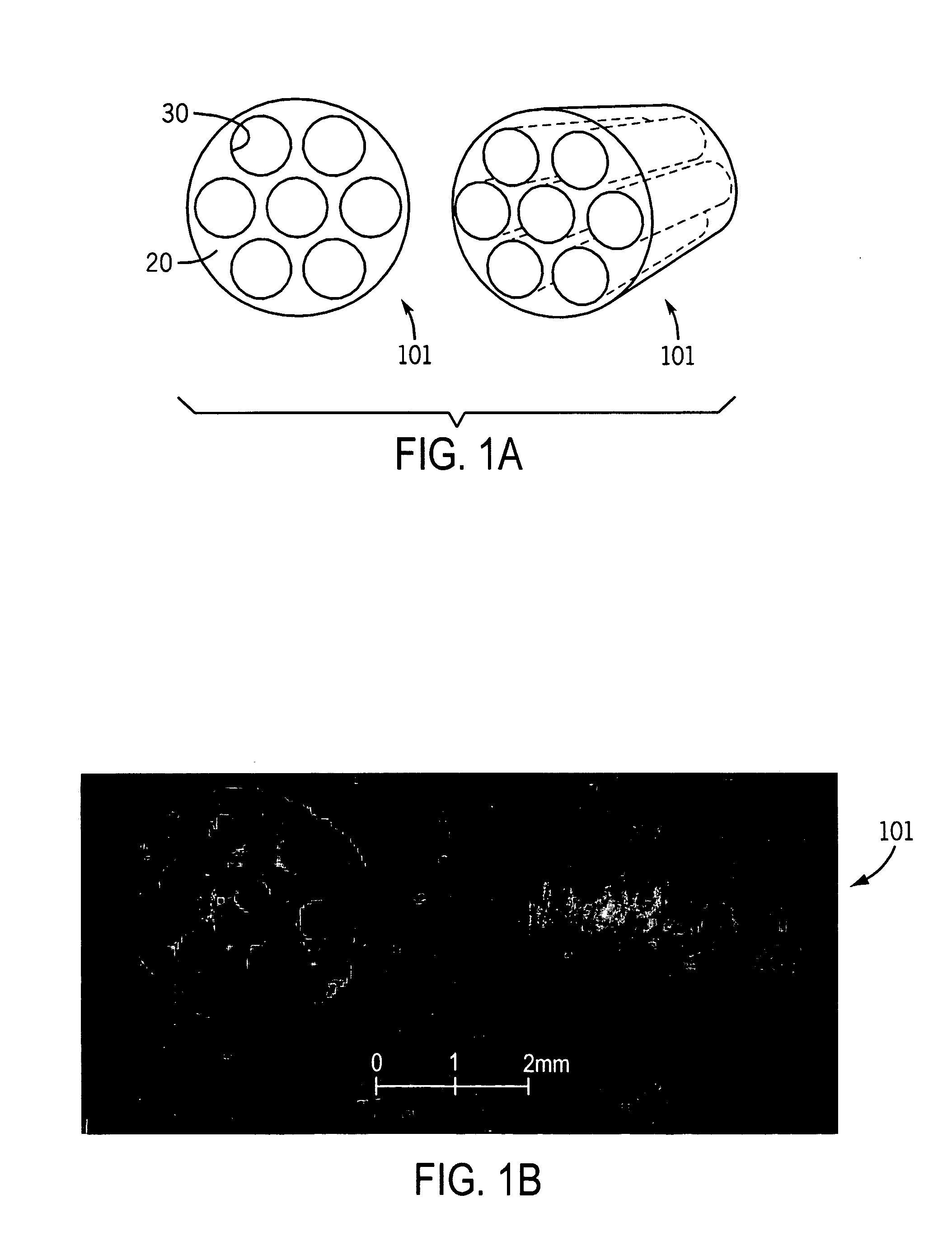
[0043]Scaffolds with parallel-channel architecture were fabricated by injecting a 50% (w / v) solution of poly-(lactic-co-glycolic acid) (PLGA), with a copolymer ratio of 85:15 lactide to glycolide, in dichloromethane (DCM) into a 3-mm diameter Teflon mold fitted with a...

example 2

Release of Chondroitinase ABC from PLGA Microparticles

2A. Introduction

[0046]Chondroitinase ABC (C-ABC) has been investigated for clinical chemonucleolysis of herniated intervertebral discs (see Olmarker et al., Spine, 1996. 21:1952–56), and recently it has been used experimentally for in vitro and in vivo degradation of proteoglycans involved in glial scarring in central (see Bradbury et al., Nature, 2002. 416:636–40) and peripheral nervous system injury (see Zuo et al., Experimental Neurology, 2002.176:221–28). Sustained, local delivery of C-ABC may therefore prove useful for multiple clinical applications. Experiments with release of C-ABC from polymer microparticles were conducted. The activity of C-ABC after release from PLGA (85-15) microparticles was investigated. Activity was examined as a decrease in absorbance, which is calculated by subtracting experimental values from blank microparticle values. The control was calculated by subtracting an aqueous positive control from th...

example 3

3A. Introduction

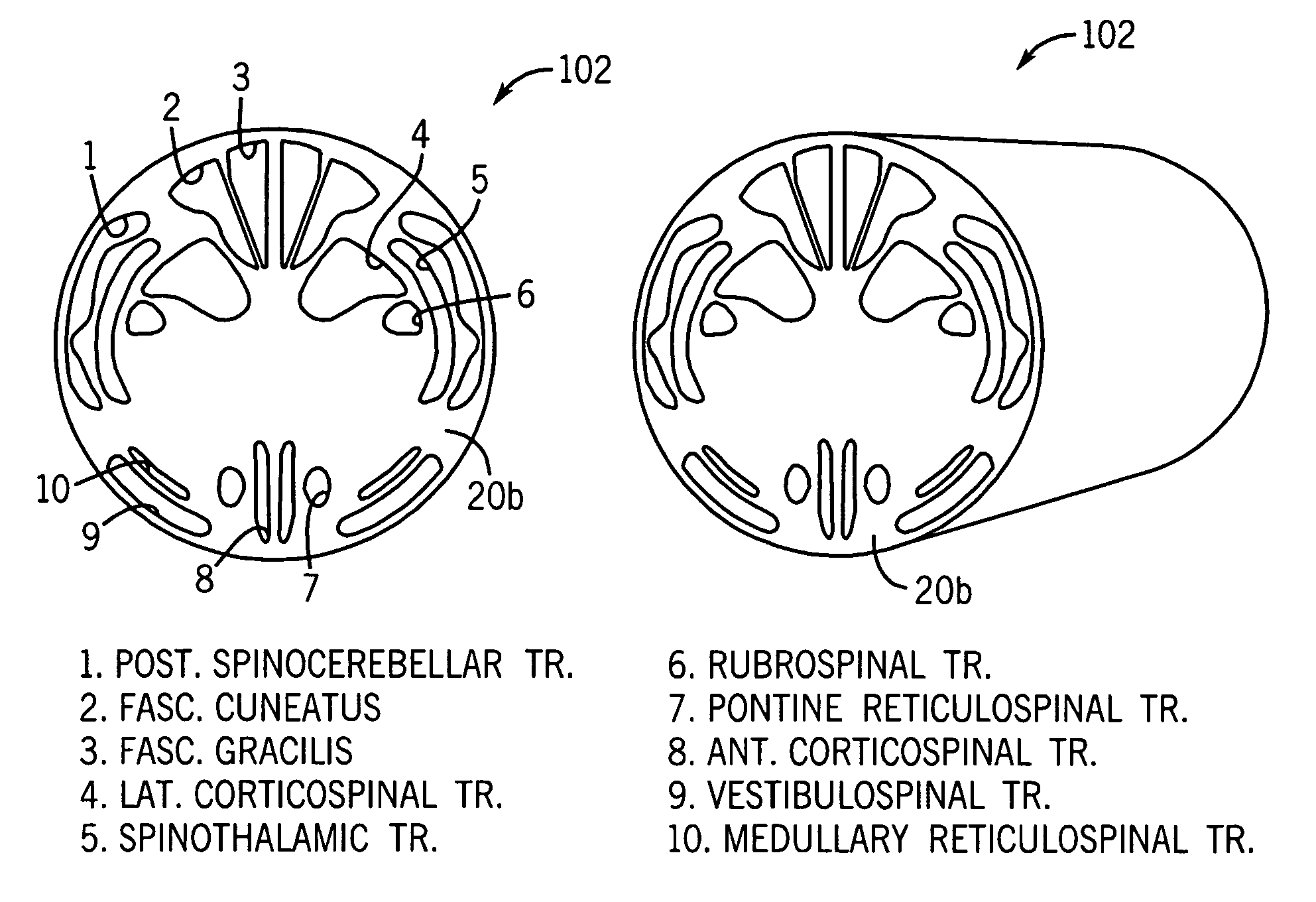
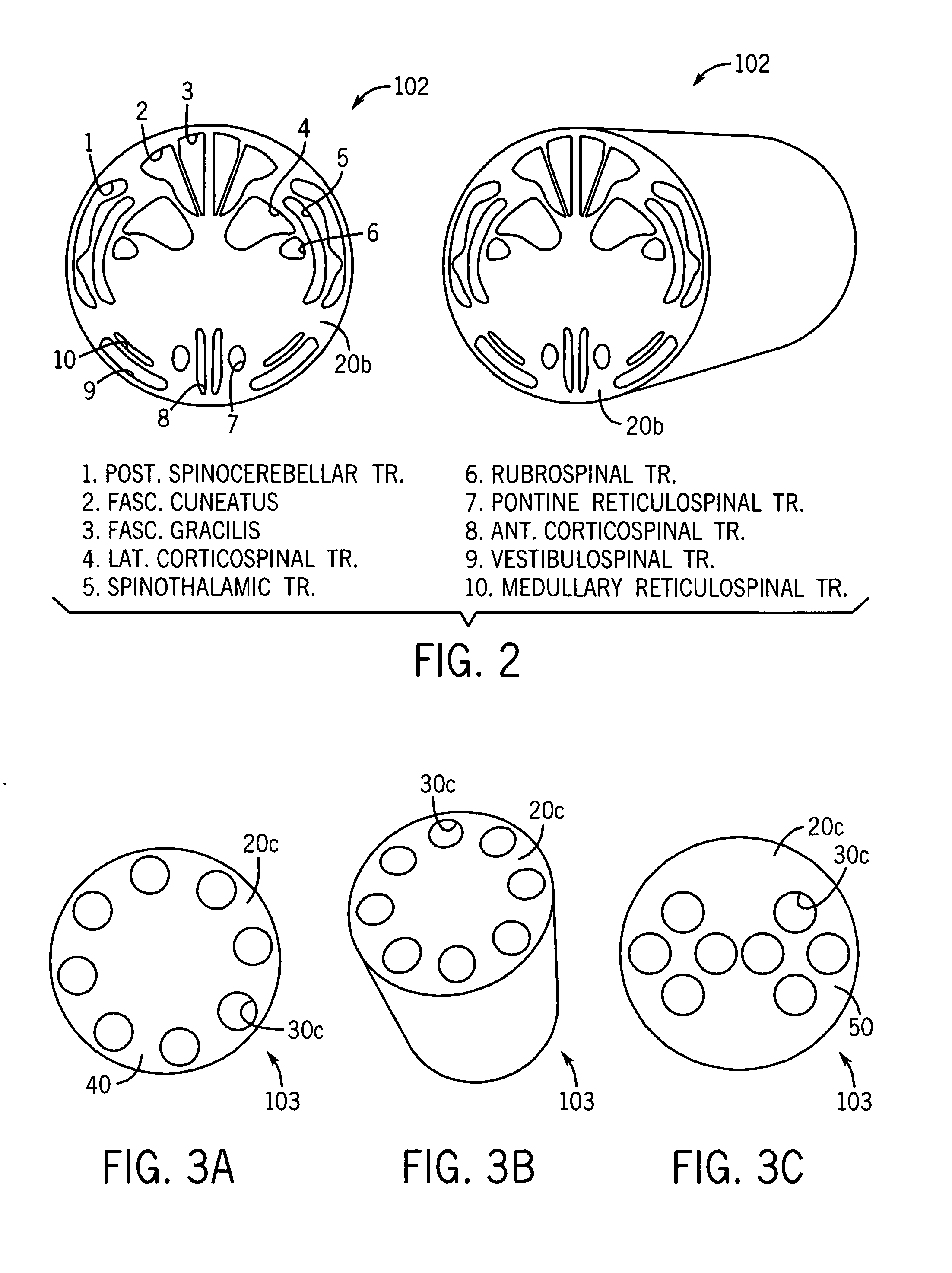
[0055]Strategies were developed to promote axonal regeneration by manipulating the structural, cellular, and molecular environment of the spinal cord. Computer aided design has been used to facilitate fabrication of grafts from the biodegradable polymer polylactide-glycolic acid. In one form, grafts were 3 millimeters in diameter and 3 millimeters in length and contained seven parallel-aligned channels of 450 micrometers diameter. Primary Schwann cells (120×106 cells / ml. in Matrigel) were microinjected into these channels. It has been demonstrated that cells survive in this environment for 48 hours in vitro. Schwann cell loaded channels were then inserted into a 3 millimeter gap between the transected ends of rat spinal cords at T8–10 level. Cords were harvested at 1, 2 and 3 months after implantation of the grafts and sectioned transverse or longitudinal to the spinal cord axis. Immunostaining against neurofilament light chain demonstrated axonal growth throughout t...


PUM


Abstract

An implant for bridging a gap in a severed spinal cord or nerve and for promoting nerve regeneration has a matrix comprising a biocompatible, biodegradable, polymeric material. The matrix has a proximal end for connection to a first end of severed spinal cord and a distal end for connection to a second end of severed spinal cord. The matrix includes internal guidance channels extending between the proximal end and the distal end to facilitate rejoining of the first end and the second end of the severed spinal cord. A bioactive agent may be dispersed in the matrix, or disposed in the channels, or included within microspheres in the channels. The channels may be arranged such that guidance channels correspond to spinal cord tracts. Alternatively, guidance channels may be spaced apart at the proximal end of the matrix and converge inward toward the axis of the matrix at the distal end.

Description

CROSS-REFERENCES TO RELATED APPLICATIONS[0001]This application claims the benefit of U.S. Provisional Patent Application No. 60 / 399,250 filed Jul. 29, 2002.STATEMENT REGARDING FEDERALLY SPONSORED RESEARCH[0002]Not Applicable.BACKGROUND OF THE INVENTION[0003]1. Field of the Invention[0004]This invention relates to a biodegradable polymer device for surgical implantation in patients with spinal cord injury.[0005]2. Description of the Related Art[0006]Despite significant advances in our understanding of neurophysiology and spinal cord injury, a patient who sustains a complete spinal cord injury has only a small hope of recovery. Loss of neurologic function following complete spinal cord injury remains a devastating condition. Research has not yet yielded an effective way to stimulate the neuronal axons to regrow to replace the injured axonal structures. Current interventions offer little hope for recovery of neurologic function in patients following complete spinal cord injury, in eith...

Claims


Application Information

Patent Timeline
IPC IPC(8): A61B17/08A61B17/11A61L27/38A61L27/58A61L31/00A61L31/14
CPCA61B17/1128A61L27/383A61L27/3878A61L31/005A61L31/148A61L27/58A61L2430/32A61B17/08
Inventor YASZEMSKI, MICHAEL J.FRIEDMAN, JONATHAN A.MOORE, MICHAEL J.WINDEBANK, ANTHONY J.
Owner MAYO FOUND FOR MEDICAL EDUCATION & RES
Why Eureka
Try Eureka