
Method for measurement of hepatitis C virus

a hepatitis c virus and detection method technology, applied in the field of hepatitis c virus detection, can solve the problems of secondary infection of blood derived components, the inability of antibody testing to determine whether the serum taken during this period is infected, and the risk of secondary infection by blood derived components, so as to reduce the detection of antigen sensitivity by interfering probe binding

Inactive Publication Date: 2008-01-08
ADVANCED LIFE SCI INST
View PDF31 Cites 8 Cited by
  • Summary
  • Abstract
  • Description
  • Claims
  • Application Information

AI Technical Summary

Benefits of technology

[0022]For the purpose of reducing secondary infection when utilizing blood and materials derived from blood, it is not necessary to distinguish between infected persons and formerly infected persons, as it is sufficient to be able to determine whether or not antibody or antigen is present. The present invention therefore provides a method o...

Problems solved by technology

Antibody testing cannot determined whether serum taken during this period is infected or not.
Therefore, there is a risk of secondary infection by the blood derived components, such as blood donation, blood components, factors from blood, contaminated specimens in the window period, because blood donor is screened by the antibody test that can not exclude such specimens.
Specifically, it is highly possible that antibodies in specimens from the subject compete with the antigen-detecting probe for the binding sites, resulting in lowering the detection of sensitivity for the antigen by interfering probe binding.
However, it is difficult to prepare probes that fulfill these conditions for molecules reported to have multiple antibody-binding sites, such as HCV core antigen.
However, since antibodies often form complexes with other biomolecules whereby they become high molecular weight entities, their separation from HCV particles is difficult by methods based on physical principles.
These methods also employ special equipment during the processing steps, which makes their application difficult for mass screening, such as blood screening.
However, it will be very difficult to separate antibodies and their complex with antigens from HCV particles, because these components precipitated in same fractions.
Moreover, HCV particles often form immune complexes between the antigens in the HCV particles and antibodies that recognize them, and it is difficult to separate only the antibodies or antigens from the immune complexes.
When antibodies against HCV are present in a specimen, there are cases where the specimen donor is currently contains HCV because of active injection of HCV, while there are other cases where the specimen does not contain HCV because of HCV elimination from body by treatment or material recovery it is difficult to discriminate these therefore cases based on the presence or absence of antibodies.
However, as explained above, antibody testing cannot determine whether serum is infected during the window period after HCV infection.
Consequently, when blood-derived substances such as transfusion and blood components, blood preparations, etc. are utilized for screening by antibody testing, there is a risk of secondary infection by specimens in the window period.
Coupling this with antigen testing is desired in order to reduce this risk, but antigen testing is still not implemented for mass screening for blood donation.
However any method have a limit of the detection sensitivity and cannot measure levels below that detection sensitivity.
Thus, no testing method exists that can discriminate with 100% accuracy.
This requirement will result in higher cost of the screening, because the number of tests performed with samples should increase than present.
As already mentioned, despite the development of antibody detecting methods and antigen detecting methods, when it is attempted to detect antigen under conditions for detecting antibodies as alluded to above, the antigen cannot be efficiently detected because of the presence of antibodies which inhibit binding of probes that detect the antigen.
Even under conditions for detection of antigen, however, the adopted methods eliminate antibodies that compete against detection of antigen, as explained above, and therefore antibodies cannot be detected.
The currently reported methods, therefore, do not allow detection of antigen and antibody by a single method.

Method used


Image

  • Method for measurement of hepatitis C virus
  • Method for measurement of hepatitis C virus

Examples


example 1

Expression of HCV-Derived Polypeptide and Purification

[0063](A) Construction of Expression Plasmid

[0064]An expression plasmid corresponding to the HCV core region was constructed in the following manner. One microgram each of DNA of plasmids pUC•C11-C21 and pUC•C10-E12 obtained by incorporating clone C11-C21 and clone C10-E12 (Japanese Unexamined Patent Publication No. 6-38765) into plasmid pUC119 were digested at 37° C. for one hour with 20 μl of a restriction enzyme reaction solution [50 mM Tris-HCl (pH 7.5), 10 mM MgCl2, 1 mM DTT, 100 mM NaCl, 15 units of EcoRI and 15 units of ClaI enzyme] and [10 mM Tris-HCl (pH 7.5), 10 mM MgCl2, 1 mM DTT, 50 mM NaCl, 15 units of ClaI and 15 units of KpnI], respectively, and this was followed by electrophoresis on 0.8% agarose gel to purify an approximately 380 bp EcoRI-ClaI fragment and an approximately 920 bp ClaI-KpnI fragment.

[0065]To these DNA fragments and a vector obtained by digesting pUC119 with EcoRI and KpnI there were added 5 μl of ...

example 2

Hybridoma Construction Method

[0076]The fused polypeptide (TrpC11) prepared by the method described above was dissolved in 6 M urea and then diluted in a 10 mM phosphate buffer solution (pH 7.3) containing 0.15 M NaCl to a final concentration of 1.0 mg / ml and mixed with an equal amount of TiterMax to make a TrpC11 suspension. The suspension was adjusted to a TrpC11 concentration of 0.01 to 0.05 mg / ml and used for intraperitoneal injection into 4- to 6-week-old BALB / c mice. After approximately 8 weeks the immunized animals were further injected through the caudal vein with a physiological saline solution prepared to a TrpC11 concentration of 0.005 to 0.03 mg / ml.

[0077]On the third day after the final booster immunization, the spleens of the immunized animals were aseptically extracted, sliced with scissors, broken into individual spleen cells using a mesh, and washed 3 times with RPMI-1640 medium. A logarithmic growth stage mouse myeloma cell line PAI which had been cultured for a few ...

example 3

Preparation of Monoclonal Antibody

[0080]The hybridomas obtained by the method described in Example 2 were transplanted into mice abdomens treated with pristane, etc., and the monoclonal antibodies that were gradually produced in the ascites were collected. The monoclonal antibodies were purified by separating the IgG fraction with a Protein A bound Sepharose column.

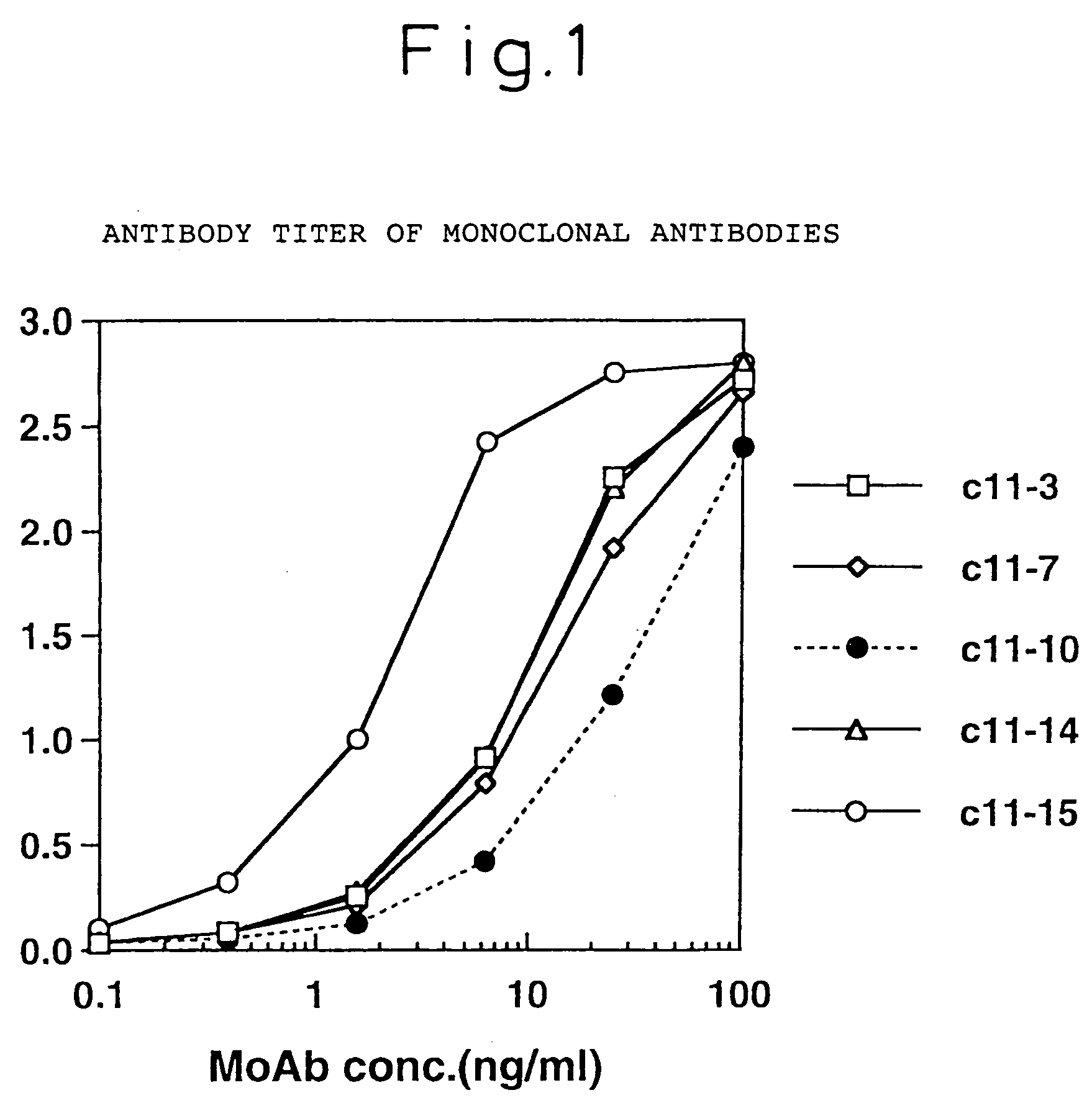
[0081]The isotype of the monoclonal antibodies produced by the above-mentioned four different hybridomas, C11-14, C11-10, C11-7 and C11-3, were identified by the double immunodiffusion method using each isotype antibody of rabbit anti-mouse IG (product of Zymed Co.), and it was found that C11-10 and C11-7 were IgG2a, and C11-14 and C11-3 were IgG1. As a result of epitope analysis of these four monoclonal antibodies using 20 peptides synthesized by HCV / core region-derived sequences, they were shown to be monoclonal antibodies which specifically recognize portions of the core sequence as shown in Table 1.

[0082]

TABLE 1Antibo...


PUM

PropertyMeasurementUnit
Fractionaaaaaaaaaa
Volumeaaaaaaaaaa
Volumeaaaaaaaaaa

Abstract

A method for measurement of the hepatitis C virus (HCV) characterized by measuring HCV core antigen and HCV core antibody by their binding with probes in the presence of an anionic surfactant or a non-ionic surfactant, or both.

Description

FIELD OF THE INVENTION[0001]The present invention relates to a method for detection of the hepatitis C virus (HCV), and more specifically it relates to a method for measurement of HCV core antigen or for simultaneous measurement of HCV core antigen and HCV core antibodies. The method is particularly effective for screening of multiple blood samples and the like.BACKGROUND ART[0002]Hepatitis caused by infection with HCV (hepatitis C virus) becomes chronic with high incidence, and as the infection period is prolonged it often progresses to liver cirrhosis and hepatocellular carcinoma. However, since infection with HCV occurs mainly through blood and blood-derived components, it is possible to identify and eliminate the source of infection to block the infection route. Current methods of identifying infection sources are primarily methods of detecting antibodies against HCV polypeptides, but methods are being sought that can identify infection sources with greater accuracy.[0003]Such m...

Claims


Application Information

Patent Timeline
IPC IPC(8): G01N33/53C12Q1/70G01N33/569C12Q1/00C07K14/00C07K14/08C07K16/00C07K16/08C07K16/10C07K2/00G01N33/576
CPCG01N33/5767G01N2333/18G01N2469/10G01N2469/20G01N33/576
Inventor AOYAGI, KATSUMIOHUE, CHIHARUIIDA, KUMIKOYAGI, SHINTARO
Owner ADVANCED LIFE SCI INST
Learn More