
Cancer immunotherapy

a technology of immunotherapy and cancer, applied in the field of cancer immunotherapy, can solve the problems of limiting the potential for autoimmune side effects that are observed, and achieve the effects of stimulating an immune response, increasing the titer of antibodies, and increasing the killing of target cells

Inactive Publication Date: 2008-03-04
PENN STATE RES FOUND
View PDF10 Cites 86 Cited by
  • Summary
  • Abstract
  • Description
  • Claims
  • Application Information

AI Technical Summary

Benefits of technology

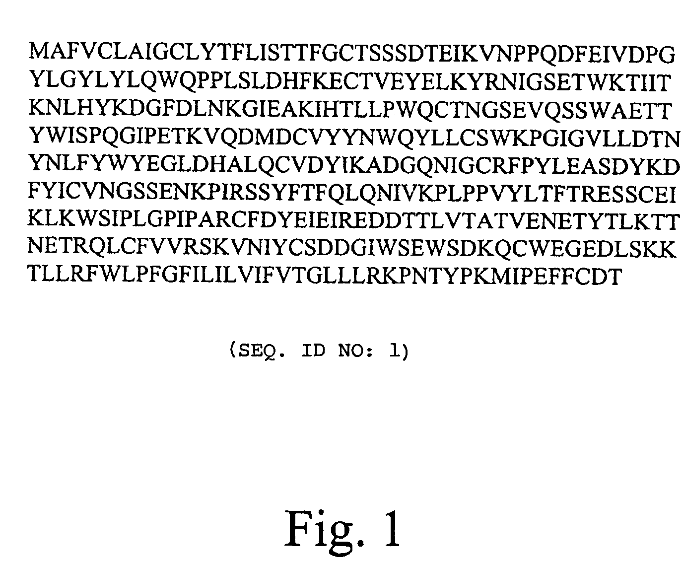
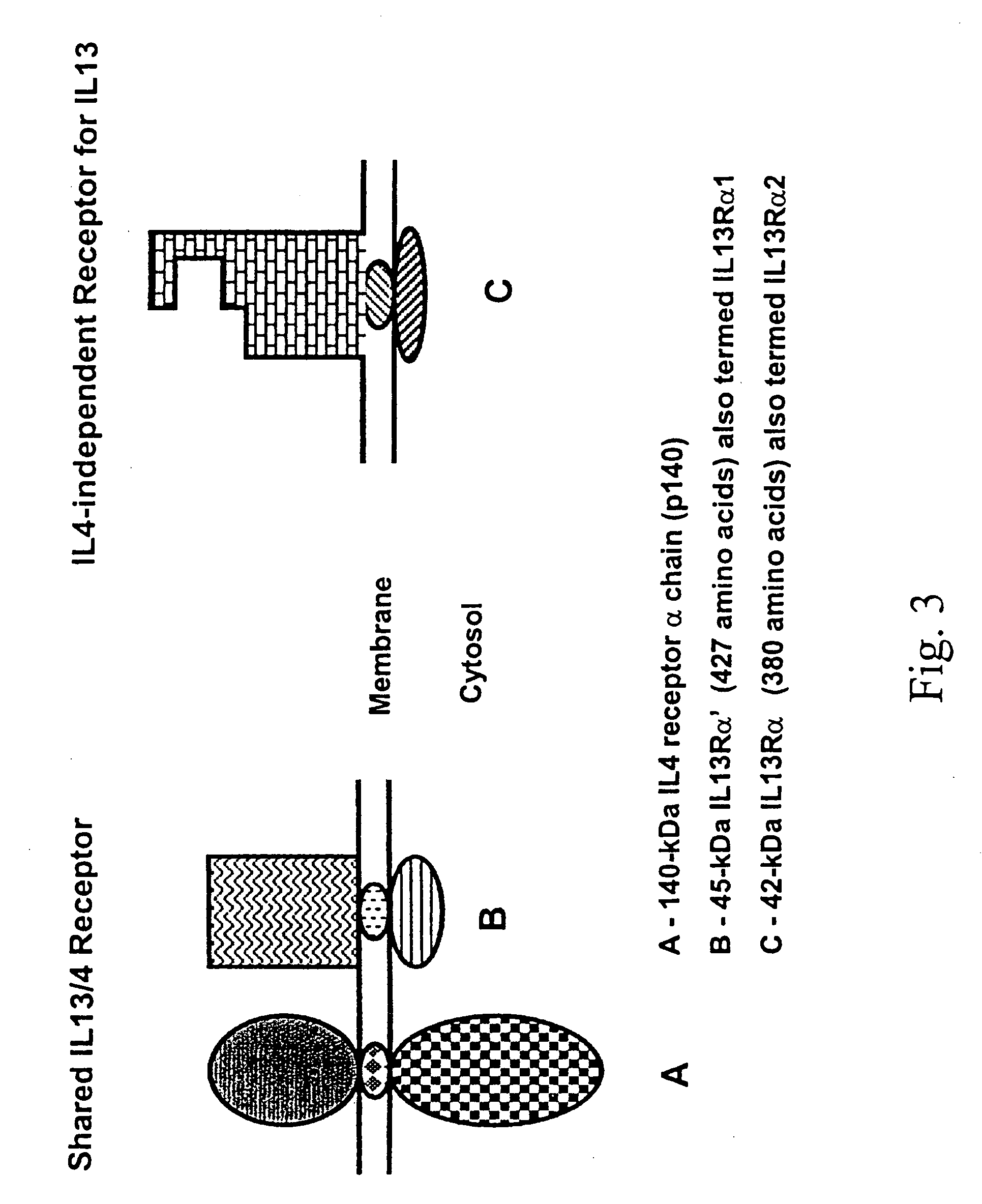
[0011]For the purpose of anti-cancer immunotherapy, IL-13Rα2 has the following distinct advantages over other cancer-related antigens. Firstly, IL-13Rα2 is a cell-surface receptor, affording it exposure to the humoral arm of the immune system. Secondly, IL-13Rα2 is expressed on the vast majority of HGGs tested, indicating its critical role in HGG progression and its potential as a target for immunotherapy. Thirdly, the physiological distribution of IL-13Rα2 is limited to cancer cells and the testes, limiting the potential for autoimmune side affects that are observed when the target is also expressed in healthy tissue. Furthermore, autoimmune side affects are unlikely because the testes are an immune-privileged organ that expresses little MHC class I molecules. Fourthly, hIL-13Rα2 is an ideal target for anti-cancer immunotherapy because of its size (380 amino acids in full length IL-13Rα2 and 343 amino acids in the extracellular domain), providing the immune system with multiple epitopes to recognize and target.
[0026]By the phrase “stimulating an immune response” is meant eliciting or increasing the activation of a lymphocyte (e.g., a B cell or T cell) or other immune system component. The stimulation of an immune response against a specific antigen can be measured as an increase in antibody titer against that antigen or the activation of one or more lymphocytes having a surface receptor specific for the antigen. Activation of lymphocytes can be determined by conventional assays, e.g., the induction of mitosis, secretion of cytokines, modulation of cell surface molecule expression, secretion of immunoglobulin (B cells), and increased killing of target cells (cytotoxic T cells).

Problems solved by technology

Thirdly, the physiological distribution of IL-13Rα2 is limited to cancer cells and the testes, limiting the potential for autoimmune side affects that are observed when the target is also expressed in healthy tissue.

Method used


Image

  • Cancer immunotherapy
  • Cancer immunotherapy
  • Cancer immunotherapy

Examples


example 1

IL-13Rα2 Mimics the Biological Features of an HGG-Associated Receptor for IL-13

[0097]Normal Chinese hamster ovary (CHO) cells were transfected with a pcDNA 3.1 plasmid (Invitrogen) containing the full length open reading frame of IL-13Rα2 and positive clones were selected with geneticin. The expression of IL-13Rα2 in these clones was tested for their ability to bind 125I-labeled IL-13. Selected clones were shown to bind labeled IL-13 independently of IL-4. In addition, labeled IL-13 was displaced by IL-13.E13K, a mutant of IL-13 shown to have a greater affinity for the IL-13 binding protein on HGG than for the shared IL-13 / IL-4 receptor found in a plethora of tissues under a physiological state. Furthermore, these IL-13Rα transfected CHO cells were exposed to an IL-13. E 13K-PE38QQR cytotoxin, a fusion protein showing potent dose dependent cytotoxicity on HGG cells. The clones expressing the receptor were killed in direct proportion to their affinity for IL-13, but not CHO cells alo...

example 2

Identification of IL-13Rα2 as a Cancer Testis Antigen

[0098]Materials and Methods

[0099]Sources of RNA. High-grade glioma cell lines A-172 MG, U-373 MG, U-251 MG and human glioblastoma multiforme explant cells (G-48) were grown in culture in appropriate media. Total RNA was extracted from the cells using the acid-guanidium isothiocyanate-phenol-chloroform method. Poly(A)+ RNA was further isolated using the Mini-oligo(dT) Cellulose Spin Column Kit (5 prime-3 prime Inc., Boulder, Colo.). 2 μg of Poly (A)+ RNA was eliectrophoresed on a 1% agarose formaldehyde gel, transferred to 0.45 μm magna charge nylon (MSI, Westborough, Mass.) and UV-crosslinked (Stratagene, La Jolla, Calif.). RNA-blotted membranes were also purchased from Clontech (Palo Alto, Calif.). Two Multiple Tissue Expresrsion (MTE™) Blots (cat # 7770-1 and 7775-1 were analyzed to determine the tissue distribution of the IL-13 binding proteins. Two sets of Human Brain Multiple Tissue Northern (MTN™) Blots (cat # 7755-1 and 776...

example 3

Representative Immunogenic Peptides of IL-13Rα2

[0113]Table I presents a list of IL-13Rα2 peptides that might be used to stimulate an immune response against IL-13Rα2 in a subject. The listed peptides were obtained using a computer program provided by the Ludwig Institute For Cancer Research (Lausanne, Switzerland). This program provided the best (at high stringency) fit of predicted immunogenic peptides that bind specific classes of MHC molecules (i.e., the various alleles of human MHC Class I indicated in Table I). The peptides indicated with the “*” are those that should bind under high stringency. The skilled artisan could produce these peptides as described herein (e.g., by automated peptide synthesis) and use each in a vaccine preparation that would be administered to a variety of test subjects (e.g. those with different MHC types) as also described herein. The immune response stimulated by each of these peptides in the subjects could then be assessed, so that those that stimul...


PUM

PropertyMeasurementUnit
Timeaaaaaaaaaa
Fractionaaaaaaaaaa
Fractionaaaaaaaaaa

Abstract

A method for stimulating a immune response against IL-13Rα2 in a subject having or at risk for developing a disease having cells expressing IL-13Rα2 includes the steps of formulating the anti-cancer vaccine outside of the subject and administering the vaccine to the subject in an amount sufficient to stimulate an immune response against IL-13Rα2 in the subject. A composition for stimulating a immune response against IL-13Rα2 in a subject having or at risk for developing a disease having cells expressing IL-13Rα2 includes an isolated agent that can stimulate immune response against IL-13α2.

Description

CROSS REFERENCE TO RELATED APPLICATION[0001]The present application is a continuation-in-part of U.S. patent application Ser. No. 09 / 780,926 filed Feb. 8, 2001 now abandoned which claims the benefit of U.S. provisional application Ser. No. 60 / 181,000 filed Feb. 8, 2000.STATEMENT AS TO FEDERALLY SPONSORED RESEARCH[0002]This invention was made with Government support under grant number CA74154 awarded by the National Cancer Institute of the National Institutes of Health. The Government may have certain rights in the invention.FIELD OF THE INVENTION[0003]The invention relates generally to the fields of biology, immunology, medicine, and oncology. More particularly, the invention relates to the use of the interleukin 13 (IL-13) receptor subunit alpha 2 (IL-13Rα2) as an immune system modulator and target for vaccines for the treatment and prevention of cancer.BACKGROUND[0004]Cancer is presently the second leading cause of death in developed nations. Wingo et al., J. Reg. Managemenit, 25:...

Claims


Application Information

Patent Timeline
IPC IPC(8): A01N37/18C12N15/09A61K35/30A61K35/76A61K39/00A61K39/39A61K39/395A61K48/00A61P25/00A61P35/00A61P43/00C07K14/715C07K16/28
CPCA61K39/0011C07K14/7155C07K16/2866A61K2039/505A61K2039/5156A61K2039/53A61K2039/545A61P25/00A61P35/00A61P35/04A61P43/00A61K39/001184
Inventor DEBINSKI, WALDEMARCHRISTENSEN, NEILMINTZ, AKIVA
Owner PENN STATE RES FOUND
Learn More