MRI contrast agents

a contrast agent and mri technology, applied in the field of in vivo reagents, can solve the problems of inapplicability of antibody-based contrast agents, almost no mri contrast agents developed for such purposes, etc., and achieve the effect of easy preparation and outstanding utility in early detection and early treatmen

Inactive Publication Date: 2008-05-06
KYUSHU UNIV
View PDF2 Cites 5 Cited by
  • Summary
  • Abstract
  • Description
  • Claims
  • Application Information

AI Technical Summary

Benefits of technology

[0007]Specifically, the present invention provides an MRI contrast agent which is novel in that, differently from the conventional MRI contrast agents which are dedicated only to imaging the overall vascular silhouette, it is capable of directly detecting exfoliated vascular endothelial sites as they are, as well as imaging the sites.
[0012]The MRI contrast agents according to the present invention are of outstanding utility in the early detection and early treatment of vascular diseases because they are capable of selectively detecting exfoliated vascular endothelia. The MRI contrast agents of the present invention can be easily prepared through known synthetic reactions and are low-cost because they are effective when used in a relatively small quantity.

Problems solved by technology

However, there are found almost no MRI contrast agents developed for such purpose.
However, such antibody-based contrast agents are not practical as they are costly because they require complicated steps to prepare and have to be used in a large amount.

Method used


Image

  • MRI contrast agents
  • MRI contrast agents
  • MRI contrast agents

Examples


example 1

Synthesis of the Contrast Agent

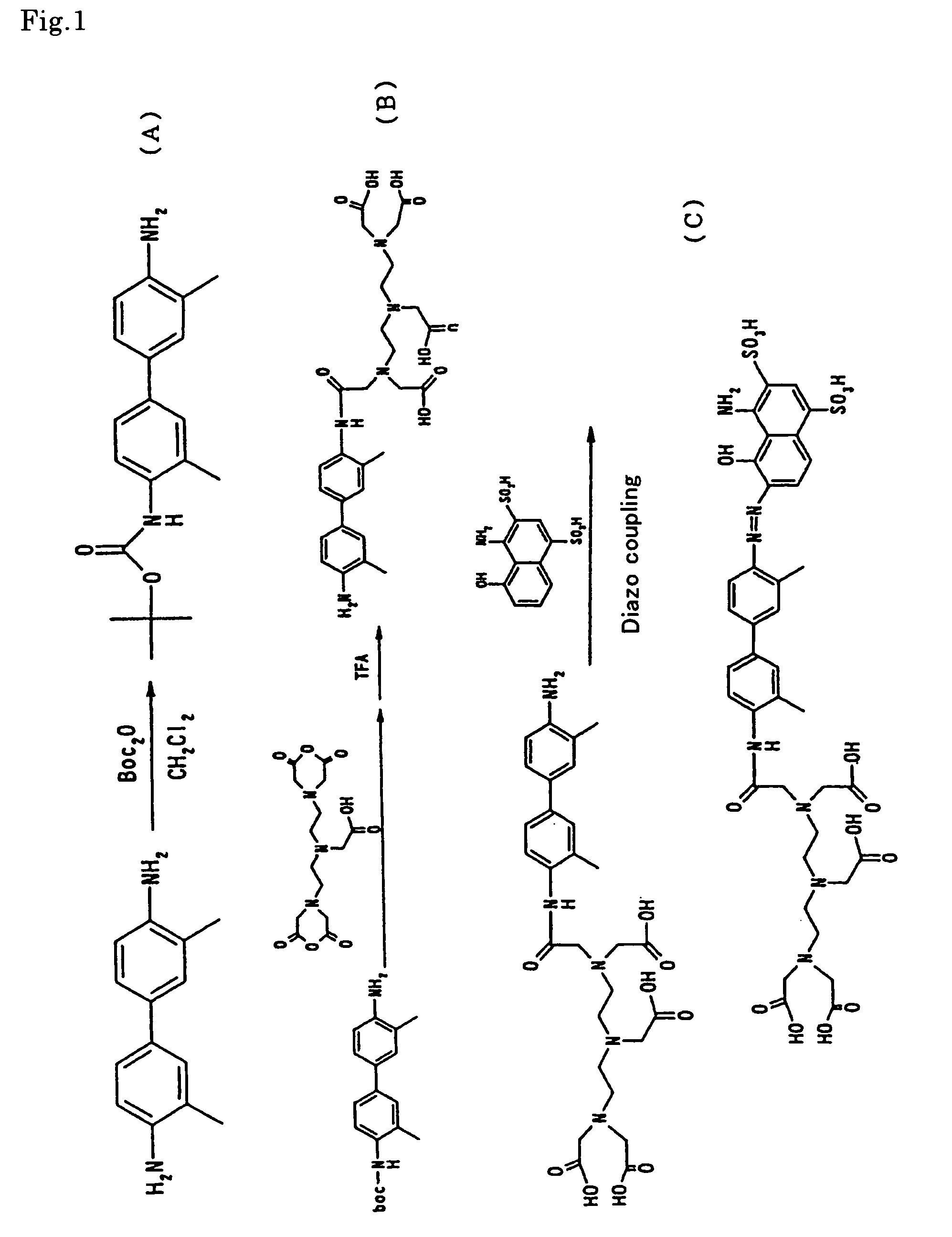
[0031]In accordance with the reaction scheme as shown in FIG. 1, there was synthesized the MRI contrast agent of the present invention as expressed by the aforementioned formula (VII).

(1) Synthesis of Boc DMB (Step A).

[0032]Dimethylbenzidine (DMB) 1.0 g (4.71 mmol), dibutoxycarbonylketone (Boc2O) 1.28 g (5.88 mmol) and triethylamine 0.714 g were added to 20 ml of dichloromethane, followed by stirring at room temperature for 48 hours. The reaction mixture was subjected to filtration, added with chloroform, and then washed with a saturated aqueous solution of L-sodium tartrate to remove unreacted DMB. The chloroform phase was subjected to concentration in vacuo, refining by silica gel chromatography, and then vacuum drying to obtain the desired product (0.60 g).

(2) Synthesis of DMB-DTPA (Step B).

[0033]Boc DMB 0.30 g (0.96 mmol) was dissolved in 20 ml of pyridine. In the resultant solution was suspended DTPA anhydride 0.343 g (0.96 mmol). The atmosphere o...

example 2

Evaluation of Imaging Capability by MRI (1)

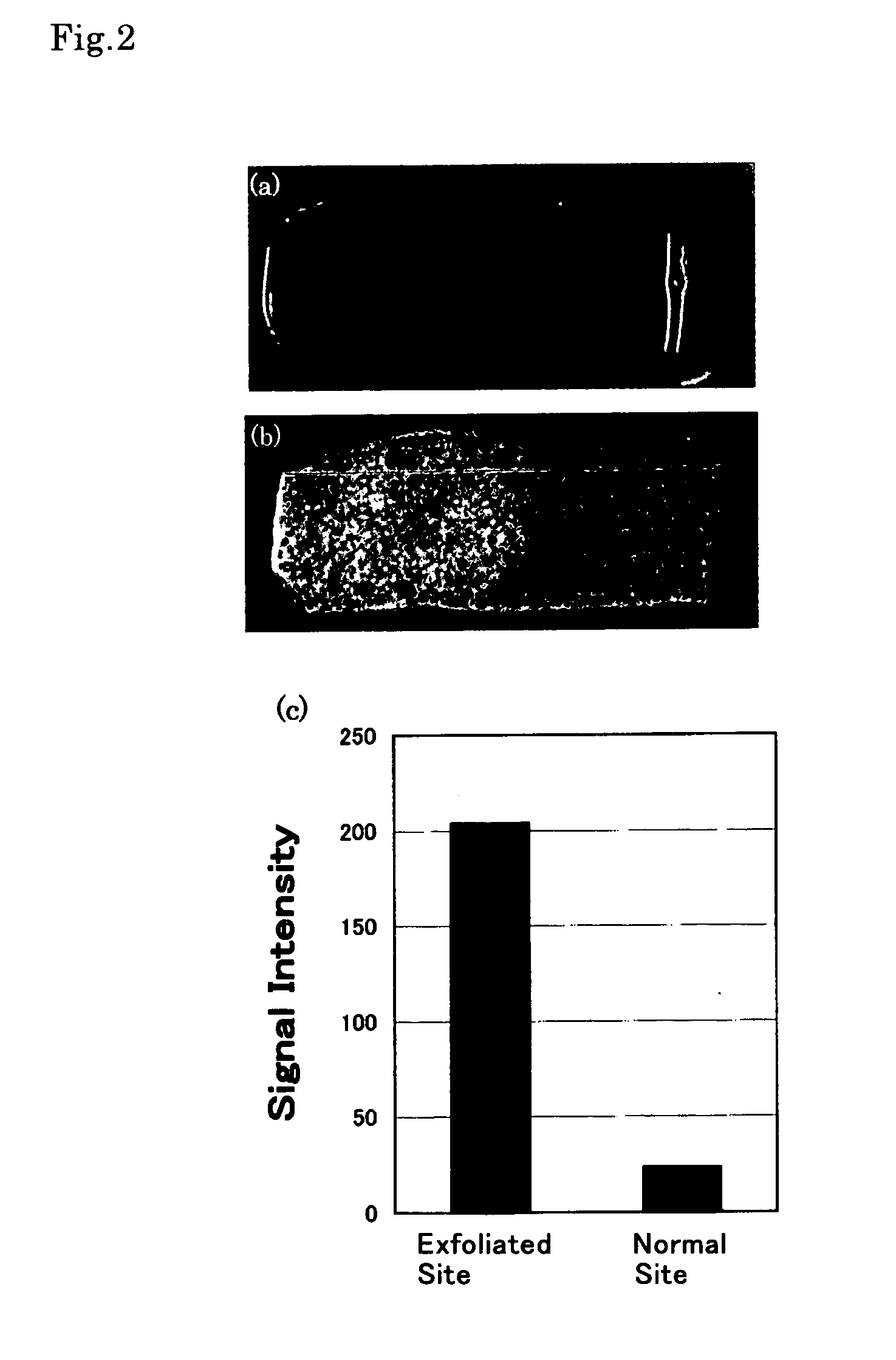
[0036]For the contrast agent as synthesized (prepared) in Example 1, evaluation of in vitro imaging capability by MRI was carried out with an aortic vascular section extirpated from a pig, as a specimen.

[0037]The aortic vascular section extirpated from a pig was spread in a rectangular shape measuring about 2 cm×3 cm to prepare a vascular section sample. The endothelial surface of the left half of the sample was exfoliated with a scalpel. The sample was then immersed in a 10 mM aqueous solution of the contrast agent for ten seconds. Following thorough washing with physiological saline solution, the sample was subjected to the evaluation by MRI as well as by visual observation.

[0038]The results are shown in FIG. 2. FIG. 2 (A) is a picture taken with a digital camera, which corresponds to the visual observation. It was visually observed that the exfoliated endothelial site (the left half) was colored blue, suggesting accumulation of the contr...

example 3

Evaluation of Imaging Capability by MRI (2)

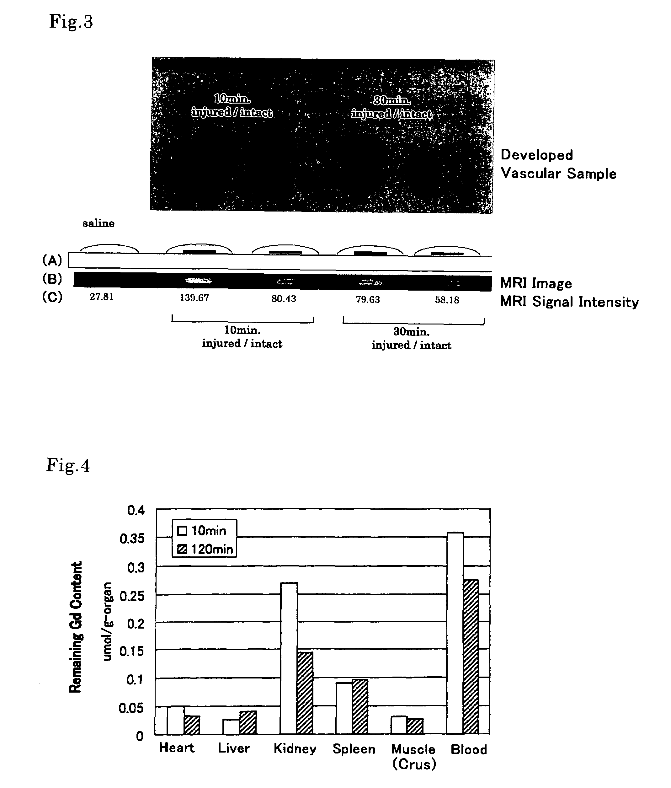
[0039]The contrast agent of the present invention was evaluated for imaging capability by means of an ex vivo experiment using a living rat.

[0040]A balloon-tip catheter was passed through the left femoral artery of the rat to injure the carotid artery with the balloon. Then 2 ml of aqueous saline solution containing 24 mM of the contrast agent (EBDTPA-Gd prepared in Example 1) was injected to the rat through the right jugular vein. After a predetermined period of time (10, 30 or 120 minutes), the right carotid artery and the left carotid artery were extirpated, developed, washed with a physiological saline, and then subjected to evaluation by MRI. The MRI evaluation was carried out by imaging the extirpated carotid artery sections, under the condition of being added with one or two droplets of physiological saline solution, by the T1 weighted spin echo method.

[0041]The results are shown in FIG. 3. In FIG. 3, by “injured” is denoted the exfo...


PUM


Abstract

An MRI contrast agent enables direct detection and imaging of exfoliated vascular endothelial sites. The MRI contrast agent includes an imaging unit which contains an unpaired electron-carrying atom and / or molecule and is capable of amplifying or reducing MRI signals, and a detection unit which is bonded to the imaging unit and is capable of selectively recognizing exfoliated vascular endothelial sites and binding thereto. The detection unit is exemplified by one having a chemical structure expressed by the following formula (I), wherein at least one of R1-R11 is, for example, a sulfonic acid group, and X is, for example, a phenyl group which may be substituted:

Description

TECHNICAL FIELD[0001]The present invention belongs to the field of in vivo reagent, and particularly relates to a novel contrast agent for use in MRI.BACKGROUND ART[0002]In MRI (Magnetic Resonance Imaging), an intravascular contrast agent is used to ensure high-contrast images. Conventional intravascular MRI contrast agents are dedicated to observation of an overall vascular silhouette, i.e., the degree of stricture in the vascular lumen to provide information for the diagnosis and treatment of vascular diseases, and they cannot be applied for directly detecting lesion sites where the vascular endothelia have exfoliated.[0003]The ability to image the exfoliated vascular endothelia directly as they are would greatly contribute to early disease detection and treatment. However, there are found almost no MRI contrast agents developed for such purpose. A method has been proposed for detecting injured or inflamed vascular sites, in which there are used antibodies against integrin or fibr...

Claims


Application Information

Patent Timeline
Patent Type & Authority Patents(United States)
IPC IPC(8): A61B5/055A61K49/00A61K49/10
CPCA61K49/085A61K49/10
Inventor KATAYAMA, YOSHIKISHIMOKAWA, HIROAKIYAMAMOTO, TATSUHIRO
Owner KYUSHU UNIV
Why Eureka
Try Eureka