Methods for the culture of human embryonic stem cells on human feeder cells

a technology of human embryonic stem cells and feeder cells, which is applied in the field of stem cell culture systems to achieve the effects of reducing the number of fibroblast feeder cells, and preventing the growth of fibroblasts

Inactive Publication Date: 2008-10-07
VIACYTE INC
View PDF12 Cites 117 Cited by
  • Summary
  • Abstract
  • Description
  • Claims
  • Application Information

AI Technical Summary

Problems solved by technology

Moreover, the potential applications for the human ES cell are limited when the ES cell is cultured in the presence of non-human feeder cell layers.
There is no evidence in the prior art showing the long-term isolation and / or maintenance of human pluripotent ES cells on non-fibroblast feeder cells.
Others have attempted to isolate human ES cells on non-fibroblast feeder cells, but have not succeeded in maintaining the human ES cells in a pluripotent state for long or indefinite periods of time.
Similarly, there is no evidence in the prior art showing the long-term isolation and or maintenance of human pluripotent ES cells in the presence of conditioned media from human cell types.
Thus the culture conditions which were successful with one large domestic animal were not successful for another domestic animal.
It is therefore not predictable that a technique successful for the isolation and short-term maintenance of ES-like cells from cows will be useful for the isolation and / or maintenance of human pluripotent stem cells.
However, none of the growth factors tested directed the differentiation of a uniform and singular cell type.
Although this procedure provided a relatively pure population, these cells cannot be used for the treatment of humans since they were cultured on mouse feeder cells.

Method used


Image

  • Methods for the culture of human embryonic stem cells on human feeder cells
  • Methods for the culture of human embryonic stem cells on human feeder cells
  • Methods for the culture of human embryonic stem cells on human feeder cells

Examples


example 1

Isolation of a Human Pluripotent ES Cell on a Mouse Embryonic Fibroblast Feeder Cell Layer

[0102]Embryo Collection: Human embryos were obtained as described by Thomson et al., 1998, supra. Briefly, fresh or frozen cleavage stage human embryos were produced by in vitro fertilization (IVF) for clinical purposes. The embryos were donated by individuals after informed consent and after institutional review board approval. The donated human embryos were cultured to the blastocyst stage in IVC-1 Medium (In Vitro Care, San Diego, Calif.) from day 0 until day 3 or day 4. After 3 days of culture the embryos were visually assessed by qualified embryologists for their quality and likely ability to develop normally. Embryos of high quality were transferred either to the patients from whom the ova were collected or were donated to other patients. The remaining embryos were cultured in IVC-3 blastocyst medium (In Vitro Care, San Diego, Calif.) and reassessed at 5 days in vitro. High quality embryo...

example 2

Characterization of a Human Pluripotent ES Cell Isolated on MEFs

[0109]The human ES cell lines that were generated in Example 1 were characterized to ensure that they were pluripotent.

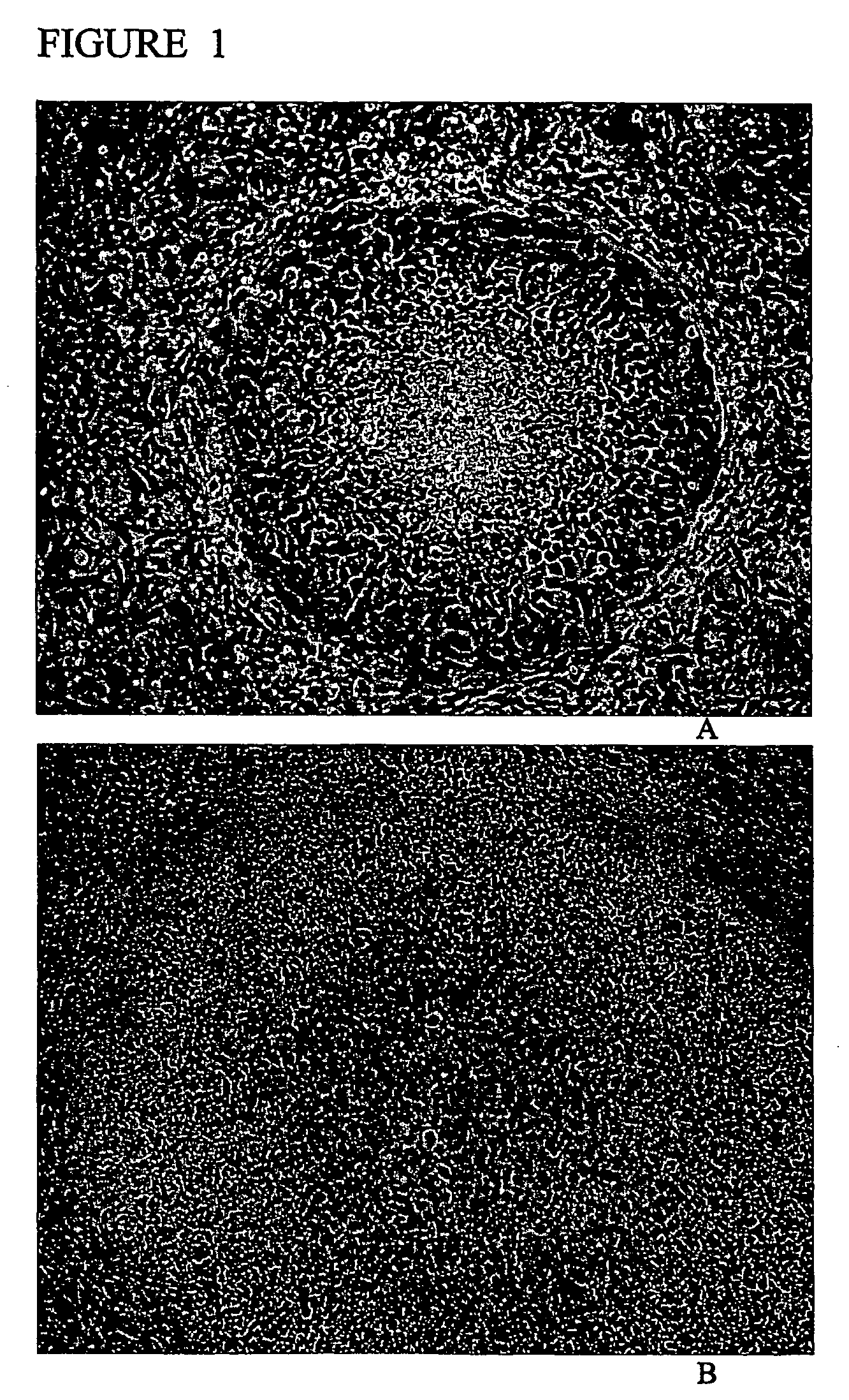
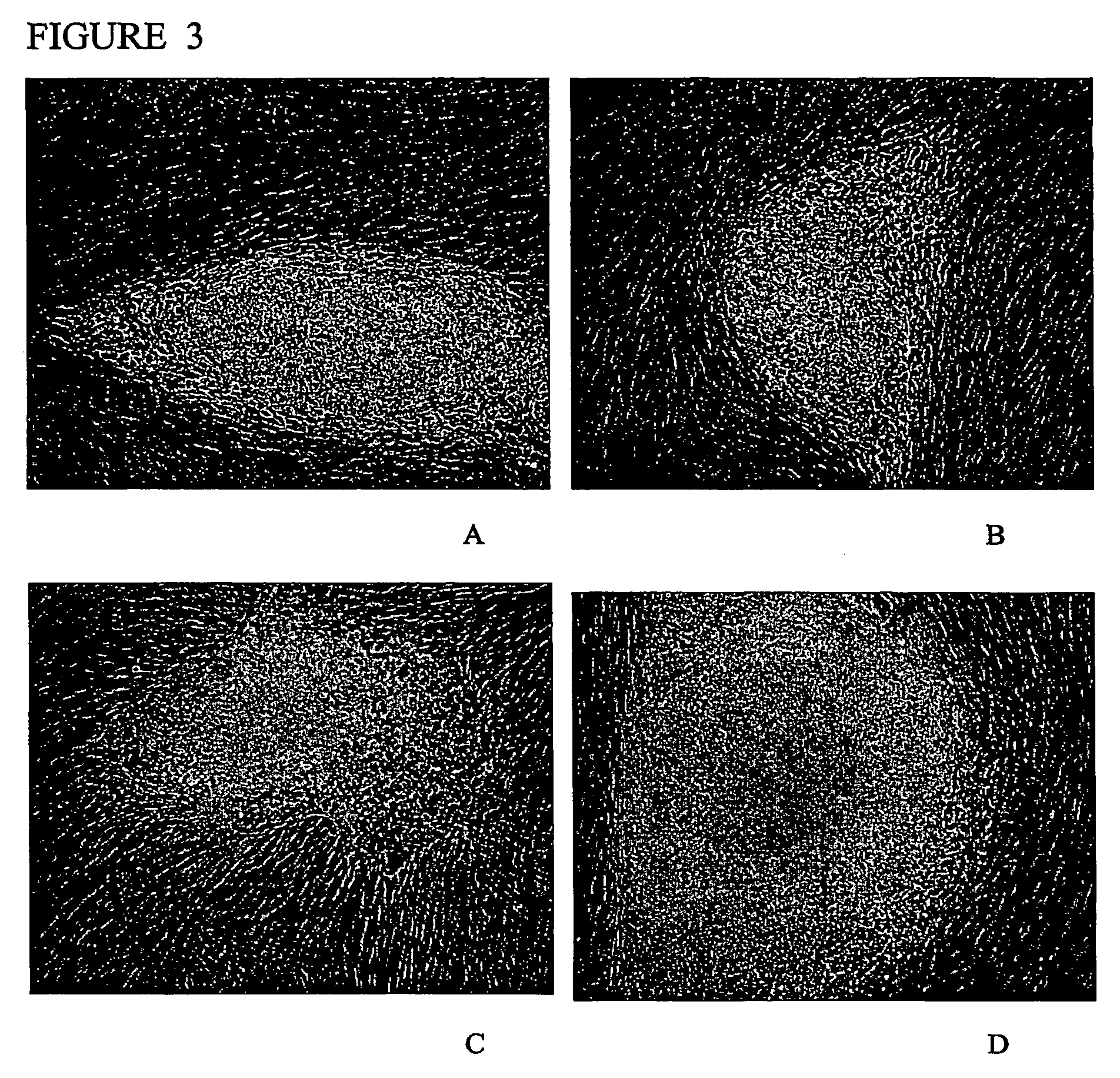
[0110]Morphology: The morphology of the derived human ES cell was similar in at least some ways to the human ES cell lines derived by other investigators (Thomson et al., 1998 supra; Reubinoff et al., 2000 supra). The human ES cells have a high nucleus / cytoplasm ratio and prominent nucleoli. Additionally, the borders between the individual ES cells are very distinguishable, particularly in comparison to mouse ES cells. The characteristic morphology of human ES cells allows for the ready identification of the cells.

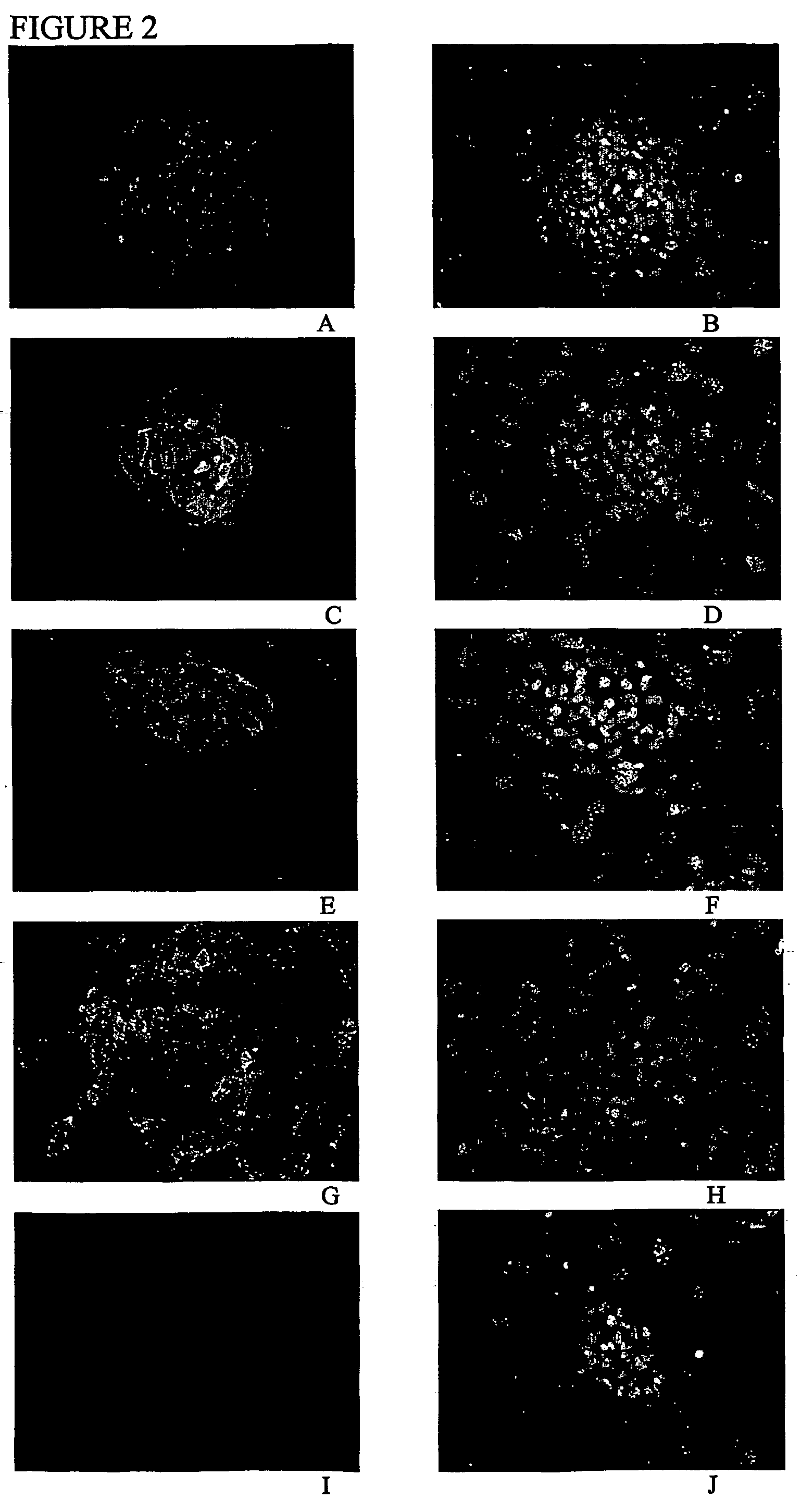
[0111]Gene expression studies: The human ES cell lines generated in Example 1 expressed alkaline phosphatase and Oct-4, two molecular markers of pluripotent cells.

[0112]Alkaline phosphatase was visualized using the Vector Red alkaline phosphatase substrate kit (Vector Lab, Burlingame, Calif.)....

example 3

Maintenance of a Human Pluripotent ES Cell on a Human Granulosa Feeder Cell Layer

[0119]Isolation of granulosa cells: Human granulosa cells were harvested at the time oocytes were collected for IVF. The oocytes were collected by laporoscopy. Oocytes have a tightly associated layer of cells surrounding them, known as the cumulus oophorus. The granulosa cells that comprise the cumulus oophorus were stripped from the oocytes by hyaluronidase treatment. The hyaluronidase treatment was followed by repeated passage of the granulosa cells through a narrow bore pipette. The granulosa cells were isolated as clumps of cells, which were further broken down to a single cell suspension by a 2 minute trypsin-EDTA treatment with 0.25% trypsin-EDTA in PBS (Gibco-BRL). After the trypsin treatment, the granulosa cells were washed with HES cell medium and plated on tissue culture dishes. The granulosa cells were cultured at 37° C. and 5% CO2.

[0120]The granulosa cells were cultured in HES medium. After ...


PUM

PropertyMeasurementUnit
timeaaaaaaaaaa
temperatureaaaaaaaaaa

Abstract

Methods and cell culture medium for the generation of human pluripotent embryonic stem cells are disclosed. Human embryonic stem cells are cultured with human granulosa feeder cells, muscle cells, Fallopian ductal epithelial cells, bone marrow stromal cells, and skin fibroblasts and the embryonic stem cells maintain their pluripotent phenotype. The human pluripotent embryonic stem cells can be cultured without feeder cells, and in the presence of supplemental growth factors. The human pluripotent embryonic stem cells can be alternatively cultured with conditioned medium obtained from a cell culture capable of maintaining human embryonic stem cells in a pluripotent state, wherein the cell culture is a human granulosa cell culture.

Description

FIELD OF THE INVENTION[0001]This invention relates generally to the isolation, maintenance, and use of stem cell cultures. Specifically, the field of the present invention is human embryonic stem cell culture system using a human feeder cell.BACKGROUND[0002]Embryonic stem cells, referred to as ES cells, are derived from the inner cell mass (ICM) of fertilized eggs in blastocyst phase, and can be cultured and maintained in vitro while being kept in an undifferentiated state. ES cells are pluripotent, possessing the capability of developing into any organ or tissue type or, at least potentially, into a complete embryo. For example, ES cells can differentiate and give rise to a succession of mature functional cells. Differentiation has been shown in tissue culture and in vivo.[0003]An important application of human ES cells is their use in cell therapy: the treatment of symptoms, diseases, conditions, and disabilities with ES cell derived replacement cells and tissues. Many diseases an...

Claims


Application Information

Patent Timeline
Patent Type & Authority Patents(United States)
IPC IPC(8): C12N5/02C12N5/08A61K35/12C12N5/0735
CPCC12N5/0606A61K35/12C12N2501/115C12N2501/235C12N2502/13C12N2502/1323C12N2502/1335C12N2502/1394C12N2502/243
Inventor MITALIPOVA, MAISAMLYONS, IAN
Owner VIACYTE INC
Why Eureka
Try Eureka