
Protein detection system

a detection system and protein technology, applied in the field of protein detection systems, can solve the problems of affecting the sensitivity, reliability, and cost of the instrument, placing substantial demands on the physical characteristics (size, weight, power consumption, etc., to achieve the effect of reducing the sensitivity (signal strength) and resolution, reducing the divergence of beams, and reducing the size of the beam

Inactive Publication Date: 2009-05-05
SANDIA NAT LAB
View PDF10 Cites 25 Cited by
  • Summary
  • Abstract
  • Description
  • Claims
  • Application Information

AI Technical Summary

Benefits of technology

[0011]The present embodiment, therefore, will provide a novel approach to attain rapid, high sensitivity analysis in a reagent-less, microfluidic bioassay.
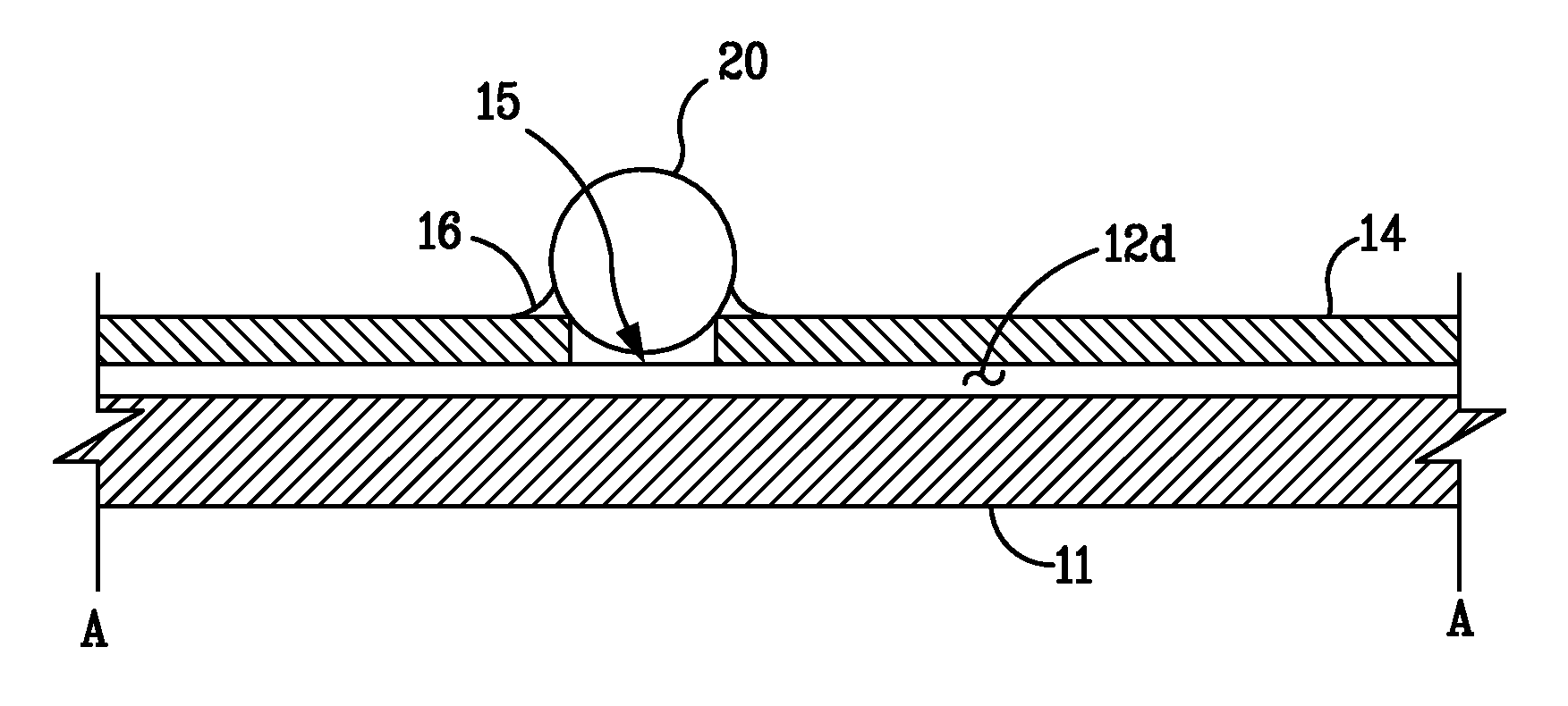
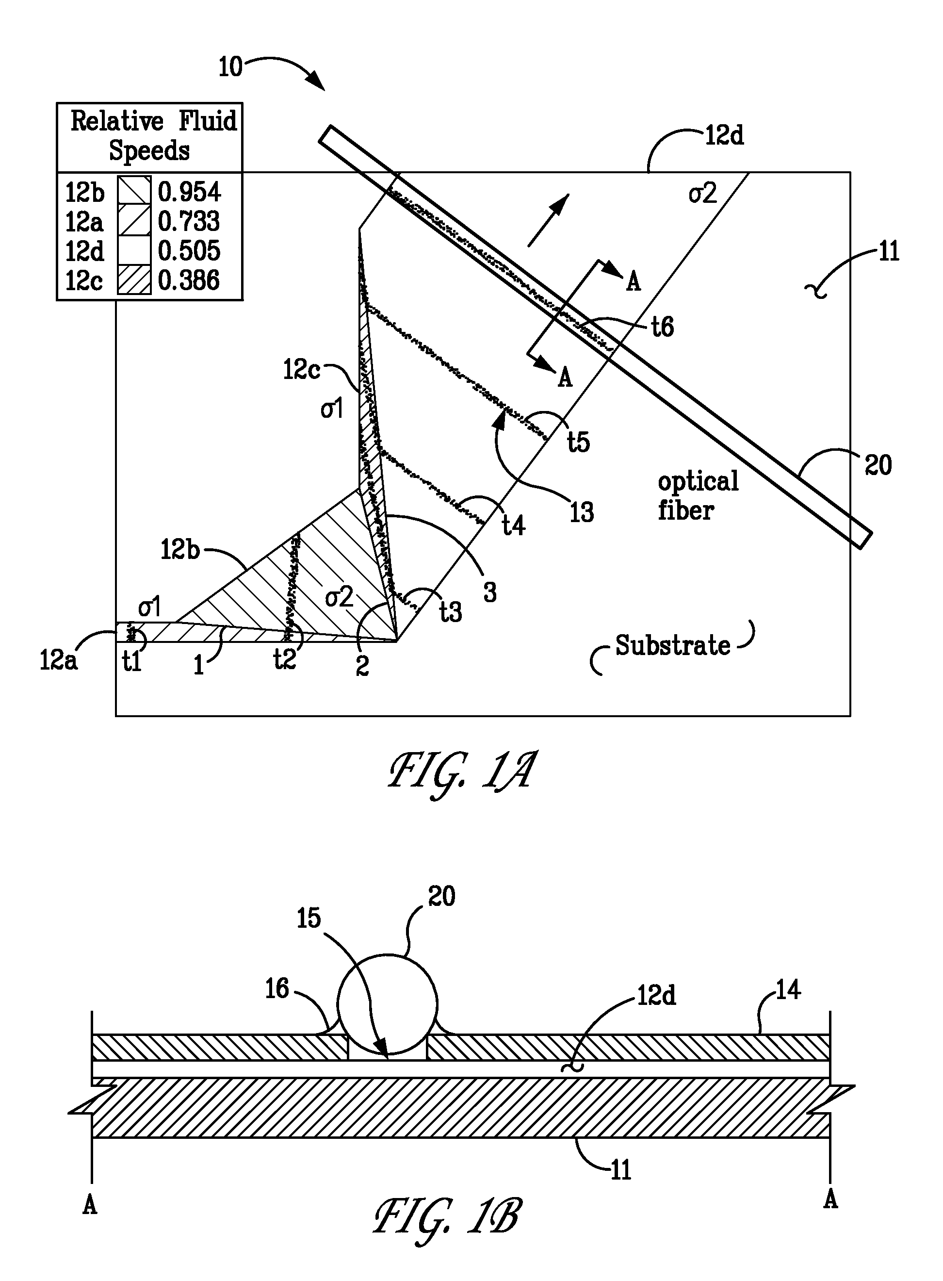
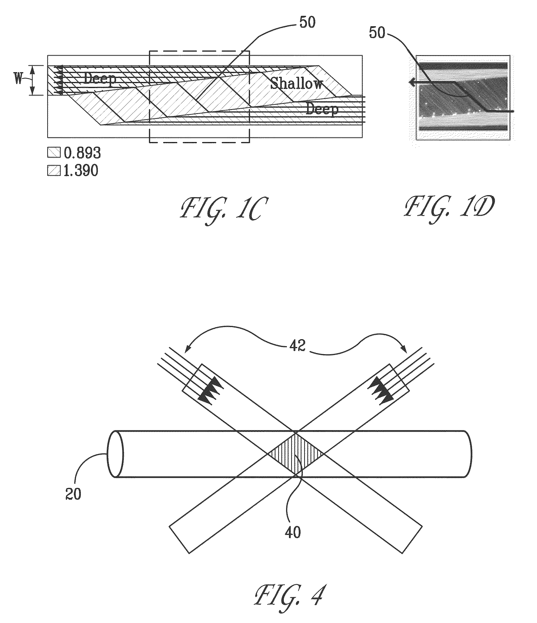
[0012]Finally, we note that these two techniques are highly complementary. For example, the evanescent field from the fiber extends into the liquid a distance on the order of the optical wavelength (˜1 μm), which is less than the depth of typical microfluidic channel. Moreover, the wide, faceted microchannels can be made shallow (also ˜1 μm in the present design), which is well matched to the fiber CRDS sensor. Similarly, the optical fiber eliminates beam divergence (diffraction), which degrades the sensitivity (signal strength) and the resolution in free-space approaches to long-path absorption.

Problems solved by technology

These applications also require high sensitivity and high specificity (i.e., low false-alarm rate), and they place substantial demands on the physical characteristics (size, weight, power consumption), reliability, and cost of the instrument.
Unfortunately, most current methods used to achieve a low detection limit rely on ultra-sensitive detection methods such as laser-induced fluorescence (“LIF”) that require significant sample processing such as fluorescent tagging (e.g., U.S. Pat. No. 6,197,503, issued Mar. 6, 2001), to be effective.
However, while a number of high-sensitivity absorption methods have been developed for gas-phase applications, condensed-phase analogs have been slow to emerge, Until recently, applications in condensed phase have been limited generally to absorption measurements of films through evanescent field experiments on the surface of all-solid state cavities and to films deposited on windows inside the cavity (see Pipino et al., Review of Scientific Instruments, 1997, v.
Unfortunately, application of absorption-based detection in microfluidic systems has been limited by poor sensitivity due to the short path lengths (e.g., 10 μm to 100 μm) characteristic of the channel width of most microfluidic devices.

Method used


Image

  • Protein detection system
  • Protein detection system
  • Protein detection system

Examples


Embodiment Construction

[0021]The low detection limits required for bio-agent sensing necessitate the use of ultra-sensitive detection methods. The most commonly used technique is fluorescent tagging followed by ultraviolet (“UV”) laser-induced fluorescence (“LIF”), which can detect protein concentrations as low as 10−12 M in microfluidic systems. Unfortunately, the time and reagents required for tagging preclude rapid detection or for long-term, unattended operation. In contrast, absorption-based methods offer the possibility of nearly universal detection without the need for labeling. Native protein detection is routinely performed in bench-top instruments using UV absorbance at 214 nm (for detecting the peptide molecule backbone) to 280 nm (for detecting protein side chains such as tryptophan). Moreover, certain specific proteins, such as myoglobin, contain chromophores that enable detection at visible wavelengths. In addition, near-infrared (“NIR”) absorbance is used to detect proteins in complex matri...


PUM


Abstract

The present embodiment describes a miniature, microfluidic, absorption-based sensor to detect proteins at sensitivities comparable to LIF but without the need for tagging. This instrument utilizes fiber-based evanescent-field cavity-ringdown spectroscopy, in combination with faceted prism microchannels. The combination of these techniques will increase the effective absorption path length by a factor of 103 to 104 (to ˜1-m), thereby providing unprecedented sensitivity using direct absorption. The coupling of high-sensitivity absorption with high-performance microfluidic separation will enable real-time sensing of biological agents in aqueous samples (including aerosol collector fluids) and will provide a general method with spectral fingerprint capability for detecting specific bio-agents.

Description

CROSS REFERENCE TO RELATED APPLICATIONS[0001]This application claims priority to prior provisional U.S. Patent Application Ser. No. 60 / 555,591 originally filed Mar. 22, 2004 entitled “PATHOGEN DETECTION SYSTEM”.STATEMENT OF GOVERNMENT INTEREST[0002]This invention was made with Government support under government contract no. DE-AC04-94AL85000 awarded by the U.S. Department of Energy to Sandia Corporation. The Government has certain rights in the invention, including a paid-up license and the right, in limited circumstances, to require the owner of any patent issuing in this invention to license others on reasonable terms.BACKGROUND[0003]Rapid detection of bio-agents is required in a number of applications, including monitoring water supplies, protecting critical facilities, process control, and hazard analysis by first responders. These applications also require high sensitivity and high specificity (i.e., low false-alarm rate), and they place substantial demands on the physical cha...

Claims


Application Information

Patent Timeline
IPC IPC(8): B01L3/02
CPCB01L3/502715G01N21/552B01L2200/10B01L2300/0645B01L2300/0654Y10T436/2575G01N21/7703G01N21/7746G01N2021/0346G01N21/359G01N2021/391
Inventor FRUETEL, JULIE A.FIECHTNER, GREGORY J.KLINER, DAHV A. V.MCILROY, ANDREW
Owner SANDIA NAT LAB
Learn More