Neural regeneration peptides and methods for their use in treatment of neural injury or degeneration

a neural injury and neurodegeneration technology, applied in the field of neurodegeneration regeneration peptides and methods for their use in the treatment of neural injury or degeneration, can solve the problems of significant neuronal cell loss and loss of brain function, no treatment available to restore neuronal function, and no treatment currently available to prevent cell death in the brain, etc., to achieve high identity, increase neuronal proliferation and migration, and induce migration, proliferation and/or neurite outgrowth

Active Publication Date: 2010-08-03
CURONZ HLDG
View PDF7 Cites 3 Cited by
  • Summary
  • Abstract
  • Description
  • Claims
  • Application Information

AI Technical Summary

Benefits of technology

[0028]Another embodiment of an NRP has been annotated, with a DNA sequence from the human genome located in the region 116668725-116667697 on the reverse strand of chromosome 6 (region according to NCBI human genome annotation project). The resulting peptide induces neuronal proliferation and migration, —as well as neurite outgrowth and survival.
[0029]Yet further embodiments of NRPs are found in rodents. One mouse NRP is encoded by a nucleic acid sequence located within the arachne contig—191157 of NCBI consisting of 339 nucleic acids using reading frame 1. Within an overlapping region, there is a second ORF of 198 nucleic acids starting at position 29 of an annotated NRP using frame 3. This ORF codes for a protein with high identity to a truncated human DNA repair protein. The resulting peptide NRP-7 induces neuronal proliferation and migration, neurite outgrowth and neuronal survival.
[0030]A still further embodiment includes NRP-8, which is also a mouse peptide, and is encoded by a nucleic acid sequence located within the genomic clone bM344E9 of the mouse Sanger database on the reverse strand. The protein coding sequence has been annotated and is located between base pairs 5609-4052. This peptide can increase neuronal proliferation and migration as well as neurite outgrowth and neuronal survival.
[0031]In another aspect, the invention includes embodiments for in vitro bioassays for evaluating proliferative and migration-inducing activity. Until recently, there were few in vitro neuronal migration assays available that could detect migrating untagged neurons over a prolonged time-period. One of these bioassays monitors olfactory peripheric placode cells organized as OTCs during a 5 day time course (Fueshko and Wray, 1994). In certain embodiments of this invention, by using in vitro bioassay using adult thalamocortical organotypic tissue cultures “OTCs,” putative NRPs can be evaluated for their ability to induce migration, proliferation, survival and / or neurite outgrowth. These embodiments can be particularly useful because 1) under control conditions, formation of a cell-bridge between both cultivated organs (e.g., thalamus and cortex) can be avoided by physically separating the two organs sufficiently far from each other (about 3 to about 5mm) on a tissue culture substrate and 2) because after birth, intrathalamic neuronal migration has been substantially completed due to the time course of thalamic ontogenesis. These bioassays can therefore be well suited for broad screening and identification of neuronal migration-inducing factors.
[0032]In certain embodiments of an in vitro assay, the thalamocortical OTC assay, includes the advantages of revealing both kind of neuronal migration, namely radial migration within the cortex and the induced tangential migration within the thalamus. Under in vitro control conditions only intrinsic cortical radial migration can be observed that due to the time course of the ontogenetic development of the neocortex.

Problems solved by technology

Moderate to severe traumatic brain injury (TBI), and focal or global ischemia can result in significant neuronal cell loss and loss of brain function within a short time period after the insult.
There are no treatments currently available to prevent cell death that occurs in the brain as a consequence of head injury or damage caused by disease.
To date, there is also no treatment available to restore neuronal function.
No drugs are currently available to intervene in the disease process or prevent cell death.

Method used


Image

  • Neural regeneration peptides and methods for their use in treatment of neural injury or degeneration
  • Neural regeneration peptides and methods for their use in treatment of neural injury or degeneration
  • Neural regeneration peptides and methods for their use in treatment of neural injury or degeneration

Examples


example 1

Identification of Human and Mouse NRPs

[0156]Using bioinformatic tools, we identified NRPs within the human, rat and mouse genomes. These new NRP genes were annotated using the following methods.

[0157]We performed a BLASTP search using the 16 amino acid rat NRP-1 as a template. We found a sequence having 100% identity to the rat NRP-1 sequence: a human cachexia-related protein. The cDNA of the human cachexia-related protein is encoded by 5 exons located on human chromosome 12. Because of the identity of the rat NRP-1 and a portion of the human cachexia protein, a new annotation of NRP orthologs was orientated alongside these 5 exons. tBlastN searches within the human NCBI-database revealed a previously unknown open reading frame (ORF) of 321 nucleic acids on chromosome 13. This sequence encodes a peptide having striking homology to the cachexia-related protein fragment having amino acids 1-30 (cachexia protein without signal sequence). These methods were also used to identify other h...

example 2

In Vitro Assay for Evaluating Migration-Inducing Activity I

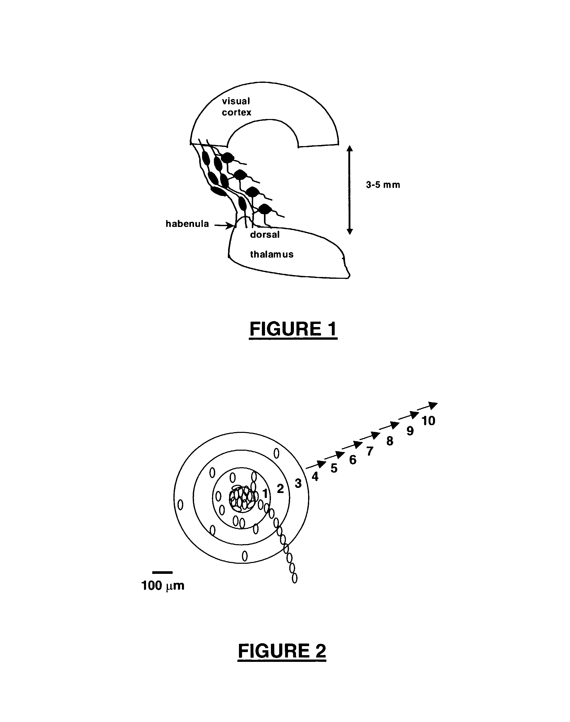
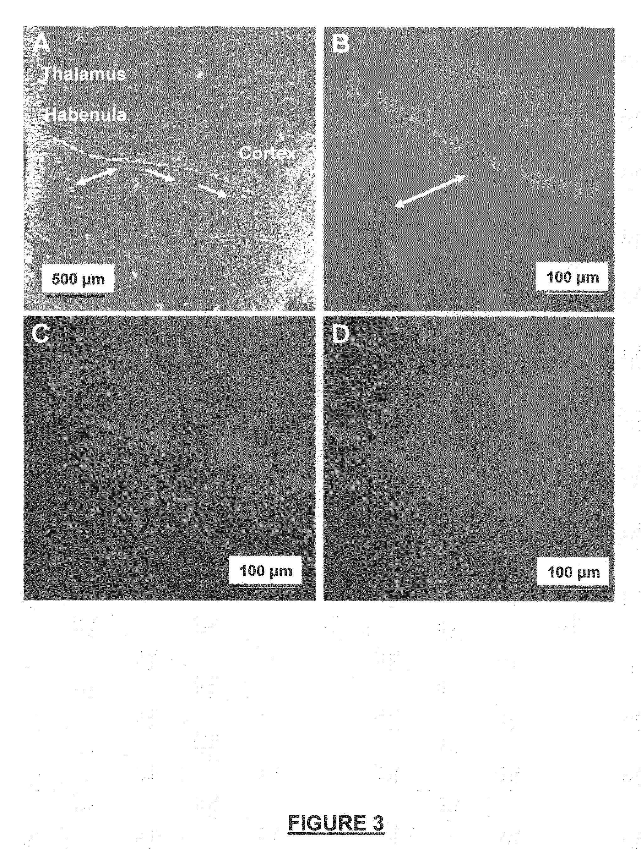
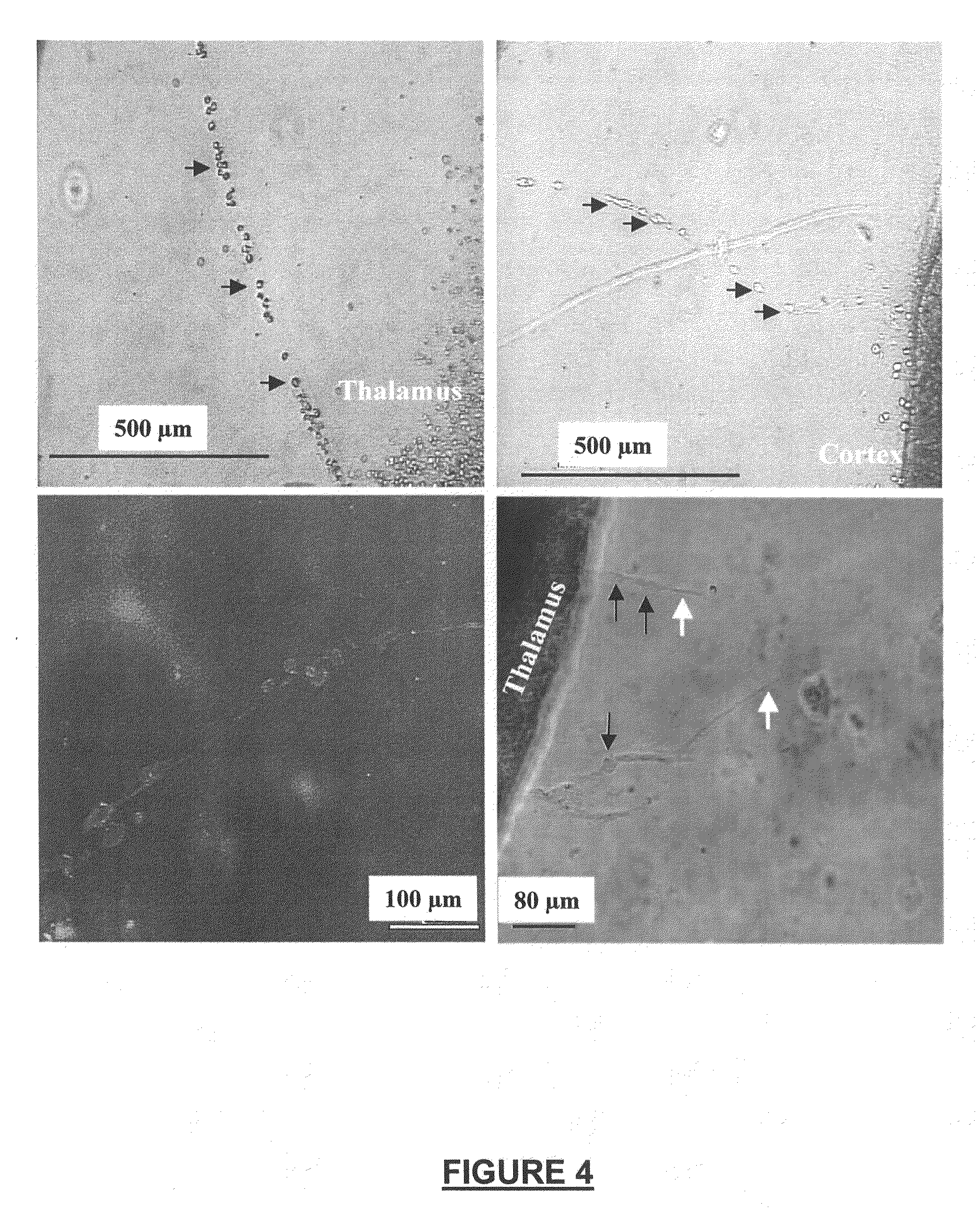
[0161]We developed a new assay system for identifying NRP having migration-inducing acgtivity. The assay system was used following guidelines approved by the Gesundheitsamt Magdeburg animal ethics committee. Newborn Long Evans rats (P0) were killed by decapitation, and neural tissues were used for preparation of organotypic cultures (OTCs). Neocortical tissue (areas 17-18 according to the Paxinos rat atlas of the developing rat brain) and thalamic tissue from the dorsal thalamus (visual areas) were extracted. These areas represent the visual axis. The dorsal thalamus was accessed by an intersection cut to remove the hypothalamus. Subsequently, the thalamus was sliced frontally using a McIllwain tissue chopper into 350 μm thick slices. Using a dissecting microscope, the habenula nucleus served as a landmark to select only dorsal thalamic areas. Cortical tissues were cut using two sagittal and two frontal intersections in orde...

example 3

Migrating Cells are of Neuronal Origin and Adopt a Differentiated Phenotype

[0169]To determine the cellular nature of cell bridges, we used neural-specific immunohistochemitsry. Immunohistochemistry was carried out according to methods of (Obst and Wahle, 1995). OTCs as described above were rinsed twice in 0.1 M phosphate buffer for 3 h. After a study was carried out, tissues were fixed using conventional fixatives suitable for immunohistochemistry. To improve antibody penetration into the tissues and cells, OTCs were incubated for 10 min in a freezing solution consisting of 25% sucrose; 10% glycerol; 100 mM NaCl in 0.01 M phosphate buffer (pH 7.4) at −80° C. (Gúlyas et al., 1996). OTCs were then incubated for 5 min in 1% H2O2 followed by a treatment of 0.4% Triton 100 and 10% normal goat serum (blocking solution) for 3 h (Sigma chemicals). Primary antibodies (anti-parvalbumin IgG; anti-calretinin IgG; anti-MAP-2 IgG) were incubated with 0.4% Triton; 2% BSA; 2% normal goat serum in P...


PUM

PropertyMeasurementUnit
timeaaaaaaaaaa
diameteraaaaaaaaaa
concentrationsaaaaaaaaaa

Abstract

The invention discloses a family of neuronal migration-inducing, proliferation-promoting and neurite outgrowth promoting factors, termed NRP compounds, and provides compositions and methods for the use of NRP compounds in the treatment of brain injury and neurodegenerative disease. NRP compounds induce neurons and neuroblasts to proliferate and migrate into areas of damage caused by acute brain injury or chronic neurodegenerative disease, such as stroke, trauma, nervous system infections, demyelinating diseases, dementias, and metabolic disorders. NRP compounds may be administered directly to a subject or to a subject's cells by a variety of means including orally, intraperitoneally, intravascularly, and directly into the nervous system of a patient.

Description

CROSS-REFERENCE TO RELATED APPLICATIONS[0001]This application claims the priority under 35 USC 119(e) to U.S. Provisional Application Ser. No. 60 / 314,952, filed Aug. 24, 2001, which is incorporated into this application fully by reference.BACKGROUND[0002]1. Field of the Invention[0003]This invention is directed to compositions and methods for the use of peptides that promote neuronal migration, proliferation, survival and / or neurite outgrowth. More specifically, this invention is directed to the use of such peptides in the treatment of brain injury and neurodegenerative disease.[0004]2. Related Art[0005]Moderate to severe traumatic brain injury (TBI), and focal or global ischemia can result in significant neuronal cell loss and loss of brain function within a short time period after the insult. There are no treatments currently available to prevent cell death that occurs in the brain as a consequence of head injury or damage caused by disease. To date, there is also no treatment ava...

Claims


Application Information

Patent Timeline
Patent Type & Authority Patents(United States)
IPC IPC(8): C07K2/00C07K7/00C07K4/00C07K14/00C12P21/00C12N15/09A61K38/00A61P9/10A61P25/00A61P25/14A61P25/16A61P25/28C07K14/47C07K14/475C12N1/15C12N1/19C12N1/21C12N5/10C12Q1/02
CPCC07K14/4756A61K38/00A61P9/10A61P25/00A61P25/14A61P25/16A61P25/28
Inventor SIEG, FRANKHUGHES, PAUL
Owner CURONZ HLDG
Why Eureka
Try Eureka