
Methods and compositions for the treatment of open and closed wound spinal cord injuries

a spinal cord injury and composition technology, applied in the direction of drug compositions, prosthesis, cardiovascular disorders, etc., can solve the problems of further damage to the spinal cord, edema of the cord itself, and damage to the vertebrae, so as to relieve the site of pressure and avoid injury

Active Publication Date: 2013-02-19
MASSACHUSETTS INST OF TECH +1
View PDF22 Cites 5 Cited by
  • Summary
  • Abstract
  • Description
  • Claims
  • Application Information

AI Technical Summary

Benefits of technology

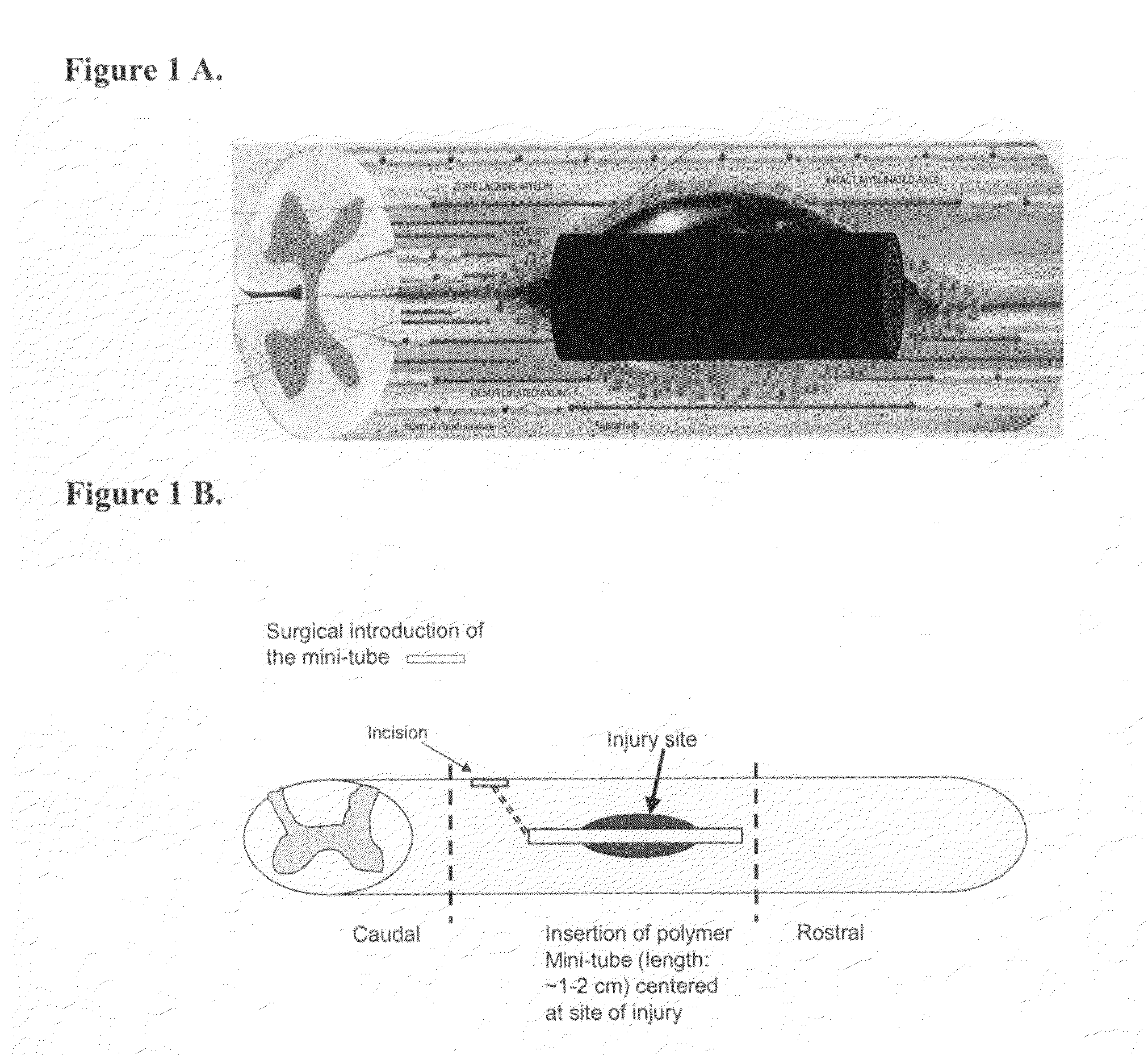
[0007]Certain embodiments of the present invention are directed to biocompatible polymeric materials which can be fabricated into “mini-tubes,” or “tubular articles.” These mini-tubes can be used to treat any localized SCI. In one embodiment, the mini-tube is inserted into the epicenter of the injury, wherein the hollow tube runs through the injury site. See FIG. 1. The mini-tube can be inserted through a surgical incision made rostral or caudal to the lesion to be treated. The mini-tube creates a new interface within the compressed spinal cord parenchyma. This new interface relieves the site of pressure and protects tissue that has been spared from injury. Pressure resulting from the compression force exerted on the cord is alleviated by (1) diffusing or redirecting the force down the surface of the mini-tube and away from the initial compressed site, and (2) absorbing the compression energy into the biocompatible material of the mini-tube. See FIG. 1. Furthermore, by providing a structure between the injured site and surrounding tissue (the new interface), inflammation may be mitigated in the adjacent area where functionally relevant residual cord tissue can be spared.
[0012]In yet another embodiment, the biocompatible polymeric materials of the present invention can be fabricated into readily formable or moldable bandages, or neuropatches. In one embodiment, a SCI is localized and the bandage or neuropatch is hand-formed to complement the injured site (for example, a hemi-sected spinal cord). The hand formed bandage is then implanted into the epicenter of the injury, wherein the bandage fills in the injury site. The implanted bandage bridges any gap formed by the spinal cord lesion and functions as an artificial pathway, nurturing regrowing neurons, reorganizing neurites and helping to form functional synapses. This new bandage interface allows for interactions between endogenous neural cells (including neural stem cells, if incorporated onto the bandage) and the inhibitory molecule-free polymer implant environment to promote cell survival. Furthermore, by providing a structure between the injured site and surrounding tissue (the new interface), inflammation may be mitigated in the adjacent area where functionally relevant residual cord tissue can be spared.
[0019]In one embodiment, the biodegradable and / or bioabsorbable polymer contains bioabsorbable and biodegradable linear aliphatic polyesters such as polyglycolide (PGA) and its random copolymer poly(glycolide-co-lactide) (PGA-co-PLA). The FDA has approved these polymers for use in surgical applications, including medical sutures. An advantage of these synthetic absorbable materials is their degradability by simple hydrolysis of the ester backbone in aqueous environments, such as body fluids. The degradation products are ultimately metabolized to carbon dioxide and water or can be excreted via the kidney. These polymers are different from cellulose based materials, which cannot be absorbed by the body.

Problems solved by technology

Insult to the spinal cord often results in vertebrae, nerve and blood vessel damage.
The pressure from the surrounding bone and meninges structure can further damage the spinal cord.
Moreover, edema of the cord itself can additionally accelerate secondary tissue loss.
In addition to damaged regeneration pathways, reflexia hyperexcitability and muscle spasticity, there are further complications of respiratory and bladder dysfunction, for example.
Over time, muscle mass is lost as a result of loss of innervations and non-use.
Even in the case of incomplete motor function loss, common problems include posture, reduced walking speed, abnormal balance and gait, and lack of sufficient weight-bearing.
However, the timing of surgical decompression has been a controversial topic.
While rat studies have shown early decompression to reduce secondary injury, the results in human clinical trials have been less than consistent.
It has been difficult to determine a time window for the effective application of surgical decompression intervention in the clinical setting.
Furthermore, there are no technologies which can be used to effectively control the increase in intra-parenchyma pressure resulting from the primary SCI.
The absence of such a technology renders surgical decompression surgery, in many cases, ineffective.
The removal of bone and soft tissue structures do not address the underlying problem of secondary intrinsic pressure at the SCI site.
Secondary injury will often impede the nerve regeneration and / or nerve regrowth process.
Reports on combat related open wound SCIs during the Vietnam war indicate that this type of injury leads to close to 100% lethality.
While there have been advances in the protective ability of bullet-proof vests, the neck region of persons wearing many of today's vests is often vulnerable to many high velocity weapons.

Method used


Image

  • Methods and compositions for the treatment of open and closed wound spinal cord injuries
  • Methods and compositions for the treatment of open and closed wound spinal cord injuries
  • Methods and compositions for the treatment of open and closed wound spinal cord injuries

Examples


example 1

Polypyrrole Mini-Tube Fabrication (I)

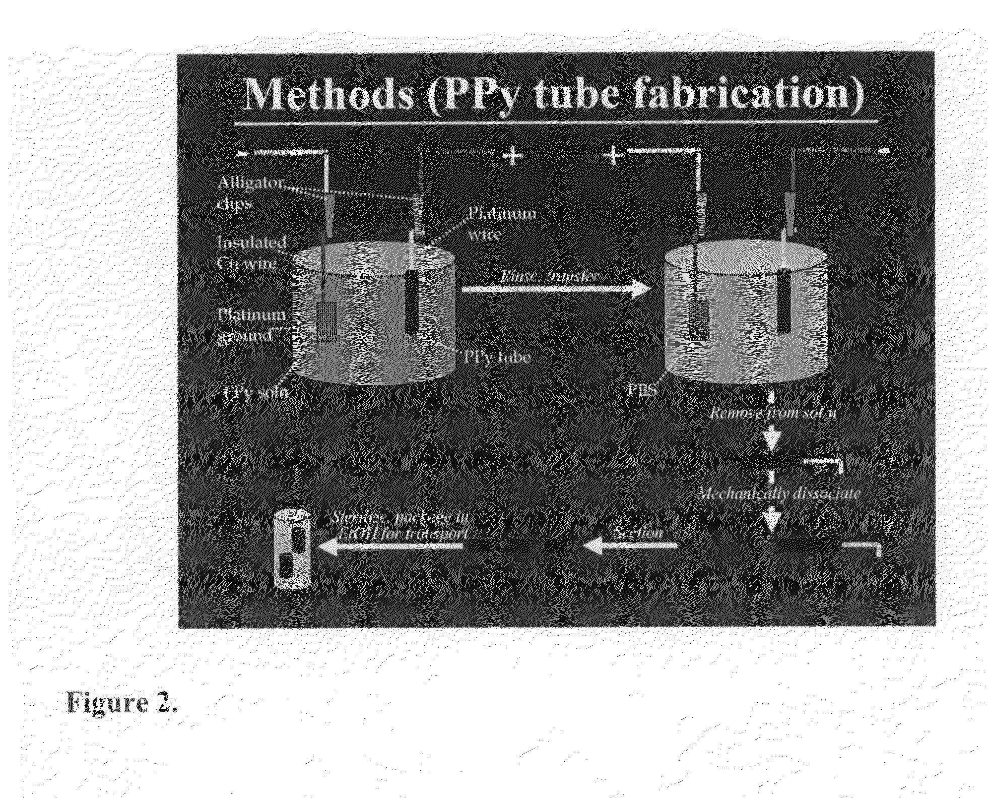
[0060]Polypyrrole tube scaffolds are created by electrodeposition of erodible PPy at 100 μA for 30 minutes onto 250 μm diameter platinum wire. See FIG. 2. This is followed by reverse plating at 3 V for 5 minutes, allowing for the removal of the scaffold. See FIG. 3 (C and D).

example 2

PPy Mini-Tubes Prevent Post-Primary Injury Cavity Formation in the Lesioned Spinal Cord (n=13, SCI and Control Rats, Respectively)

[0061]MRI images of post-injury cavity development, studied two months post injury, show large cavity formation in the control spinal cord (wherein injured cord was not treated with surgically implanted mini-tube), as compared to the PPy-treated spinal cord. See FIG. 4.

example 3

Open-Field Locomotor Scores for Polypyrrole-Implanted Rats and Lesion Control Rats

[0062]Results from the polypyrrole mini-tube scaffold showed functional locomotor improvement as early as 2 weeks post injury. The amount of functional recovery relative to non-treated controls continues to increase for up to 6 weeks. See FIG. 5. Treated animals are capable of weight-bearing and functional stepping, where non-treated animals show greatly diminished hindlimb function. Magnetic resonance images show that the fluid filled cyst is reduced with a herein described implant. As shown in FIG. 4, the spinal cord is more intact and the cyst is barely visible when treated with polypyrrole. Biodegradable and / or biocompatible polymers are well known in the art and can be used in the present invention.


PUM

PropertyMeasurementUnit
diameteraaaaaaaaaa
diameteraaaaaaaaaa
diameteraaaaaaaaaa

Abstract

Devices and methods for the treatment of open and closed wound spinal cord injuries are disclosed. For example, described herein are devices and methods for mitigating secondary injury to, and promoting recovery of, spinal cord primary injuries. More particularly, certain embodiments of the present invention are directed to polymeric mini-tubes that may be used for the treatment of spinal cord injuries. In addition, other embodiments are directed to polymeric “fill-in” bandages that may be used for the treatment of spinal cord injuries. For example, an erodible, or biodegradable, form of biocompatible polymer of the present invention is fabricated for surgical implantation into the site of the spinal cord injury.

Description

RELATED APPLICATION[0001]This application claims the benefit of U.S. Patent Application Ser. No. 60 / 794,986, filed on Apr. 25, 2006, the contents of which are incorporated herein by reference in their entirety.BACKGROUND OF THE INVENTION[0002]Many spinal cord injuries (SCIs) are a result of the spinal cord being compressed, not cut. Insult to the spinal cord often results in vertebrae, nerve and blood vessel damage. Bleeding, fluid accumulation, and swelling can occur inside the spinal cord or outside the spinal cord but within the vertebral canal. The pressure from the surrounding bone and meninges structure can further damage the spinal cord. Moreover, edema of the cord itself can additionally accelerate secondary tissue loss. There is considerable evidence that the primary mechanical injury initiates a cascade of secondary injury mechanisms including excessive excitatory neurotransmitter accumulation; edema formation; electrolyte shifts, including increased intracellular calcium;...

Claims


Application Information

Patent Timeline
Patent Type & Authority Patents(United States)
IPC IPC(8): A61F2/00
CPCA61F2/02A61L31/14A61L31/148A61L27/26A61L27/58A61L31/06A61L31/16A61F2210/0004A61L2300/412A61L2430/38A61L27/18A61L27/3834A61L27/56A61L31/005A61L31/146A61P19/00A61P25/00A61P9/14C08L67/04A61K35/12A61L27/38A61L27/54A61F2/442A61K9/0085A61L2430/00A61L31/048A61L2300/41A61L2300/414A61L2300/60A61L2300/64A61L2300/802A61L27/383A61L27/3878A61L27/50A61L2300/606A61L2400/00
Inventor LANGER, ROBERTSAIGAL, RAJIVTENG, YANG
Owner MASSACHUSETTS INST OF TECH
Learn More