
Micro-RNAs that control myosin expression and myofiber identity

a micro-rna and myofiber technology, applied in the field of development biology and molecular biology, can solve the problems of dilated cardiomyopathy (dcm), a major health risk, and a sudden death of patients, and achieve the effect of inhibiting expression or activity

Inactive Publication Date: 2013-07-09
BOARD OF RGT THE UNIV OF TEXAS SYST
View PDF62 Cites 10 Cited by
  • Summary
  • Abstract
  • Description
  • Claims
  • Application Information

AI Technical Summary

Benefits of technology

This approach effectively reduces cardiac hypertrophy and heart failure symptoms, improves cardiac function, and treats musculoskeletal disorders by modulating miRNA activity, offering a safer and more effective alternative to existing therapies.

Problems solved by technology

Heart disease and its manifestations, including coronary artery disease, myocardial infarction, congestive heart failure and cardiac hypertrophy, clearly presents a major health risk in the United States today.
The cost to diagnose, treat and support patients suffering from these diseases is well into the billions of dollars.
Scar tissue is not contractile, fails to contribute to cardiac function, and often plays a detrimental role in heart function by expanding during cardiac contraction, or by increasing the size and effective radius of the ventricle, for example, becoming hypertrophic.
While the hypertrophic response is initially a compensatory mechanism that augments cardiac output, sustained hypertrophy can lead to dilated cardiomyopathy (DCM), heart failure, and sudden death.
The causes and effects of cardiac hypertrophy have been extensively documented, but the underlying molecular mechanisms have not been elucidated.
Unfortunately, many of these standard therapies have numerous adverse effects and are contraindicated in some patients.
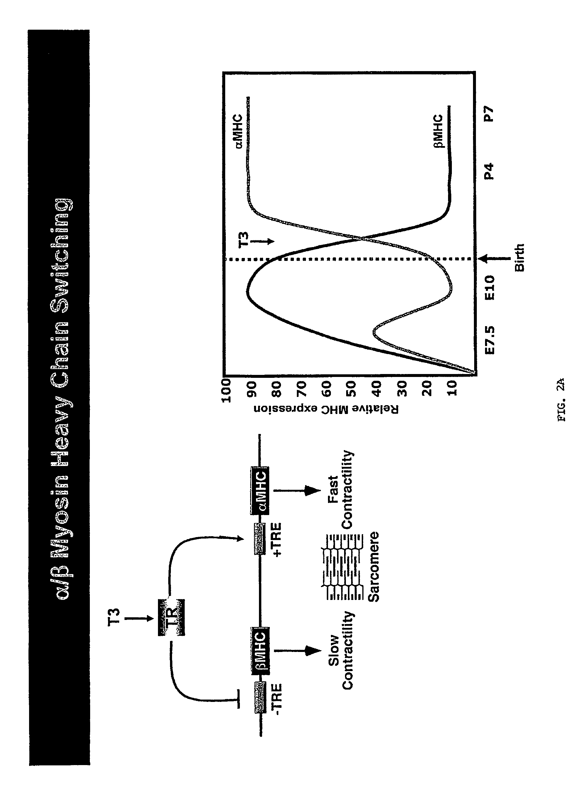
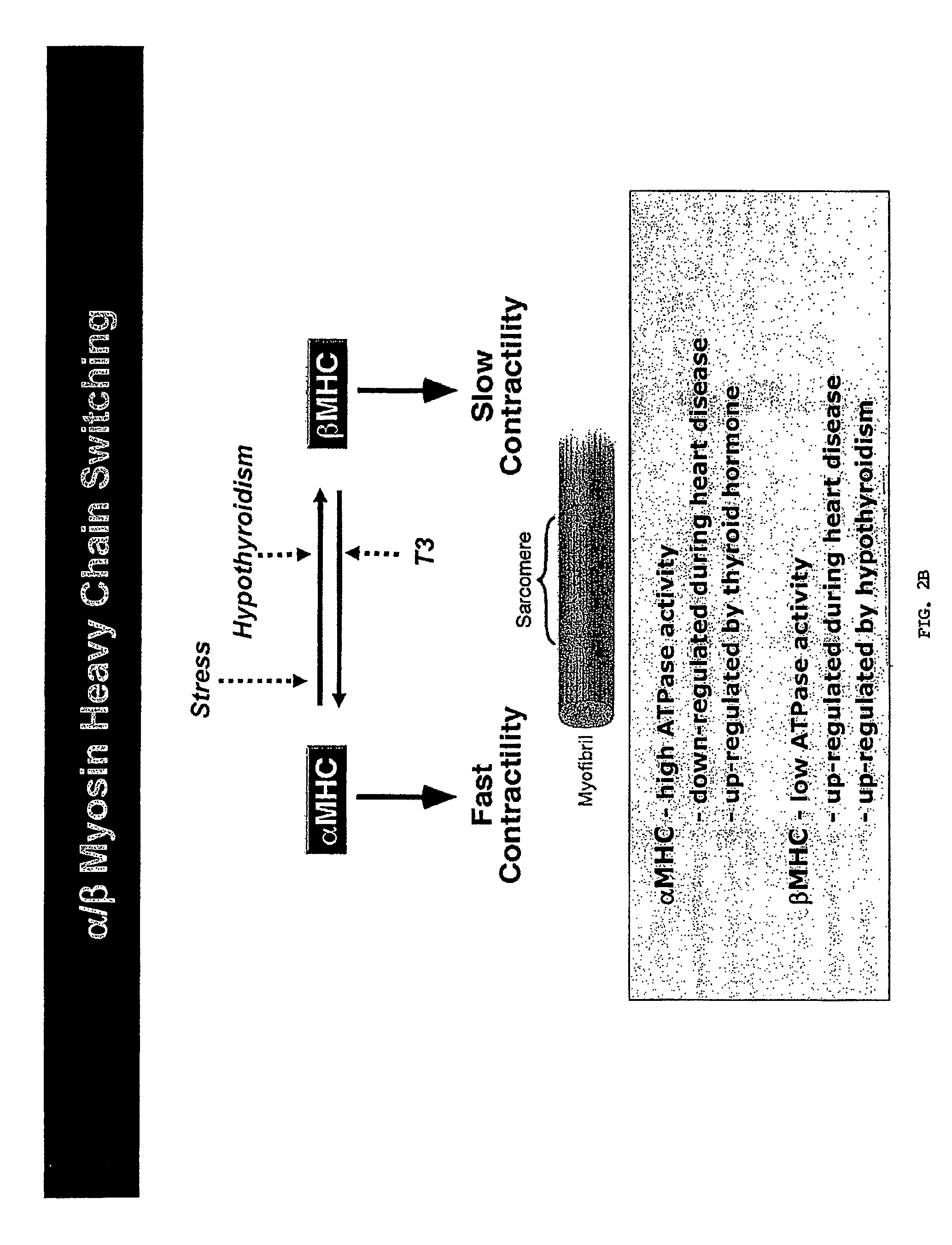
In response to a variety of pathological stimuli such as myocardial infarction, hypertension, and other disorders, β-MHC expression increases, while α-MHC expression decreases with a consequent reduction in myofibrillar ATPase activity and reduced shortening velocity of cardiac myofibers, leading to eventual contractile dysfunction.
Stress-mediated myocardial hypertrophy is a complex phenomenon associated with numerous adverse consequences with distinct molecular and histological characteristics causing the heart to fibrose, dilate and decompensate which, through myocyte degeneration and death, often culminates in heart failure.
Disruptions in myofiber electrical activity, including denervation, also lead to muscle atrophy.
Extreme disuse of a muscle, however, may result in a permanent loss of skeletal muscle fibers and the replacement of those fibers by connective tissue.
Although increasingly sophisticated computational approaches to predict miRNAs and their targets are becoming available, target prediction remains a major challenge and requires experimental validation.
Ascribing the functions of miRNAs to the regulation of specific mRNA targets is further complicated by the ability of individual miRNAs to base pair with hundreds of potential high and low affinity mRNA targets and by the targeting of multiple miRNAs to individual mRNAs.

Method used


Image

  • Micro-RNAs that control myosin expression and myofiber identity
  • Micro-RNAs that control myosin expression and myofiber identity
  • Micro-RNAs that control myosin expression and myofiber identity

Examples


example 1

miR-208 is Required for Expression of miR-499

[0268]To further explore the mechanism of action of miR-208 in the heart, the inventors defined the microRNA expression patterns in hearts from wild type and miR-208 null mice by microarray analysis. Among several microRNAs that were up- and down-regulated in mutant hearts, the inventors discovered that miR-499 was highly abundant in normal hearts, but was not expressed above background levels in miR-208 mutants. These findings were confirmed by Northern blot (FIG. 14). Analysis of the genomic location of the miR-499 gene showed it to be contained within the 20th intron of the Myh7b gene, a homolog of the α-MHC gene (FIG. 15). miR-208 appears to regulate Myh7b and thereby miR-499 expression at the level of transcription since RT-PCR for myh7b indicates that the mRNA of the host gene is dose-dependently abrogated in the absence of miR-208 (FIG. 14). The Myh7b gene is conserved in vertebrates and is expressed solely in the heart and slow sk...

example 2

MEF2 Regulates miR-499 Expression in Cardiac and Skeletal Muscle

[0269]Within the 5′ flanking region of the Myh7 gene, the inventors identified a potential MEF2 consensus sequence that was conserved across species. This sequence bound MEF2 avidly in gel mobility shift assays (FIG. 37A), and mutation of this sequence abolished both binding (FIG. 37A) and transcriptional activation of a luciferase reporter by MEF2 (FIG. 37B). The promoter region of the Myh7 gene was fused to a lacZ reporter and transgenic mice were generated. As shown in FIG. 37C, this genomic region was sufficient to direct lacZ expression specifically in the heart at E12.5. In the postnatal heart, lacZ staining was observed only in the ventricles, consistent with in situ hybridization (data not shown). Mutation of the MEF2 site completely eliminated expression of the lacZ transgene (FIG. 37C). Northern blot analysis on in vivo mouse models also showed the expression of miR-499 to be sensitive to MEF2. Cardiac-specifi...

example 3

Identification of Targets for miR-499

[0271]Given the sequence homology between miR-208 and miR-499 and the inventors' previous data demonstrating that genetic disruption of miR-208 lead to a strong induction of specifically fast skeletal muscle genes and repression of β-MHC in the heart, it is likely that miR-499 has a comparable function in skeletal muscle and could act as a dominant regulator of fiber type. Expression of miR-499 and its host transcript are regulated by the myogenic transcription factor MEF2, a positive regulator of slow fiber gene expression and muscle endurance. The inventors suggest that the fiber type regulation of miR-499 is likely to be dependent on target genes of miR-499 involved in fiber type regulation.

[0272]MiR-208 is highly homologous to miR-499 and, the remarkable fact that both microRNAs are encoded by introns of Mhc genes, suggests that they share common regulatory mechanisms. Since miRNAs negatively influence gene expression in a sequence specific m...


PUM


Abstract

The present invention relates to the identification of two microRNAs, miR-499 and miR-208b, that repress fast skeletal muscle contractile protein genes. Expression of miR-499 and / or miR-208b can be used to repress fast fiber genes and activate slow fiber genes in the treatment of musculoskeletal disorders. Inhibition of miR-499 and / or miR-208b is proposed as a treatment for cardiac hypertrophy, myocardial infarction, and / or heart failure. Pharmaceutical compositions comprising antagonists and agonists of miR-499 and miR-208b function are also disclosed.

Description

CROSS-REFERENCE TO RELATED APPLICATIONS[0001]This application is a national stage application of International Application No. PCT / US2008 / 071837, filed Jul. 31, 2008, which claims the benefit of U.S. Provisional Application No. 60 / 952,911, filed Jul. 31, 2007; U.S. Provisional Application No. 60 / 980,113, filed Oct. 15, 2007, and U.S. Provisional Application No. 60 / 980,314, filed Oct. 16, 2007, all of which are herein incorporated by reference in their entireties.STATEMENT OF GOVERNMENT SUPPORT[0002]This invention was made with grant support under grant no. HL53351-06 from the National Institutes of Health. The government has certain rights in the invention.DESCRIPTION OF THE TEXT FILE SUBMITTED ELECTRONICALLY[0003]The contents of the text file submitted electronically herewith are incorporated herein by reference in their entirety: A computer readable format copy of the Sequence Listing (filename: MIRG—004—02US_SeqList_ST25.txt, date recorded: Jun. 8, 2010, file size 9 kilobytes).FI...

Claims


Application Information

Patent Timeline
Patent Type & Authority Patents(United States)
IPC IPC(8): C12N15/11C07H21/04
CPCA61K31/7105C12N15/113C12N2330/10C12N2310/141C12N2310/113A61P21/00A61P43/00A61P9/00A61P9/04A61P9/10
Inventor OLSON, ERICVAN ROOIJ, EVA
Owner BOARD OF RGT THE UNIV OF TEXAS SYST
Learn More