Cartilage cell epimatrix three-dimensional porous sponge stent for tissue engineering and preparation method thereof

A three-dimensional porous, chondrocyte technology, applied in bone implants, medical science, prostheses, etc., can solve the problem of low biomechanical strength of cell-particle complexes, difficulty in sending metabolites from nutrients, and uneven mixing of chondrocytes To achieve the effect of extensive material sources, no risk of disease transmission, and good clinical application prospects

Active Publication Date: 2009-08-05
GENERAL HOSPITAL OF PLA
View PDF0 Cites 41 Cited by
  • Summary
  • Abstract
  • Description
  • Claims
  • Application Information

AI Technical Summary

Problems solved by technology

Preparation and preliminary study of natural biological scaffold materials for auricular cartilage. Chinese Journal of Otology, 2005, 3: 194-199), but the formed decellularized cartilage does not have a tissue engineering scaffold, which requires a porous network structure, the delivery of nutrients and It is difficult to dispatch metabolites; in addition, there are domestic research reports on decellularization of cartilage after crushing. Han Xuefeng et al. crushed cartilage into 100-154 μm particles and mixed them with chondrocytes to construct tissue engineered bone. Chondrocytes are unevenly mixed and do not have a specific shape, the biomechanical strength of the cell-particle complex is low, and surgical implantation is not easy to operate, etc.

Method used


Image

  • Cartilage cell epimatrix three-dimensional porous sponge stent for tissue engineering and preparation method thereof
  • Cartilage cell epimatrix three-dimensional porous sponge stent for tissue engineering and preparation method thereof
  • Cartilage cell epimatrix three-dimensional porous sponge stent for tissue engineering and preparation method thereof

Examples


Embodiment 1

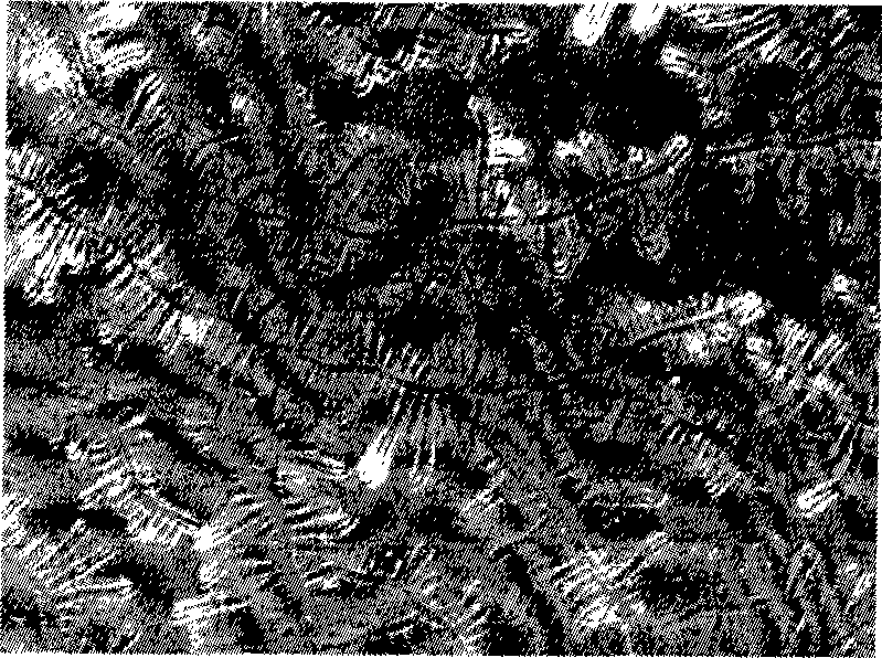
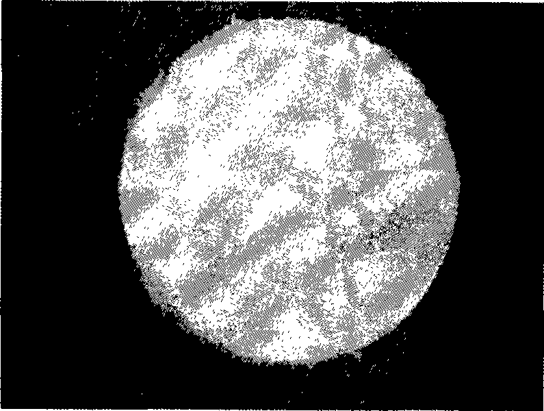
[0070] Soak human articular cartilage in PBS buffer (pH7.5) at 4°C, the buffer contains 0.035% PMSF (w / v) and 0.1% EDTA, immediately pulverize it with a pulverizer, and use gradient centrifugation to obtain cartilage with a diameter of 100nm to 5μm Microfilament ( figure 1 ). Add 10mmol / L Tris-HCl buffer solution (pH7.5) containing 1% Triton X-100 at 4°C, and the content of PMSF in the buffer solution is the same as above. Continue shaking for 24 hours to remove cellular components. Wash with PBS after centrifugation at 7000r / min for 5min. Add 50U / mL DNase and 1U / mL RNase to digest at 37°C for 6-48h. Rinse with triple distilled water after centrifuging in the same way, and the precipitate is made into a white milky suspension with a mass volume ratio of 3% ( figure 2). Put the suspension into a mold, store at -20°C for 24 hours, store at -80°C for 1 hour, and freeze-dry in a freeze dryer for 48 hours to make a three-dimensional porous scaffold material. The obtained car...

Embodiment 2

[0081] Example 2: The effect of the content of decellularized cartilage microfilaments on the microstructure of the cartilage extracellular matrix three-dimensional porous sponge scaffold

[0082] Grind the fresh articular cartilage in a pulverizer, centrifuge at 1500rpm for 5min with density gradient centrifugation, take the milk-like supernatant, use 1% Triton X-100 to continuously shake at 4°C for 48h to remove cell components, centrifuge at 7000rpm and wash with distilled water Rinse repeatedly. The precipitates were respectively made into (by weight) 1% and 2% suspensions, and the solutions were injected into pre-cooled molds, and the freeze-freeze method was used to freeze at -20°C for 24 hours, and then freeze in a freeze dryer. After drying, three-dimensional porous sponge scaffolds of cartilage extracellular matrix with different pore sizes and microstructures were obtained. see Figure 21 with 22 . Under the same freezing temperature and freezing rate, the pore s...

Embodiment 3

[0083] Example 3: Crosslinking of Cartilage Extracellular Matrix Three-dimensional Porous Sponge Scaffold

[0084] Grind the fresh articular cartilage in a pulverizer, centrifuge at 1500rpm for 5min by differential centrifugation, take the milk-like supernatant, use 1% Triton X-100 to shake continuously at 4°C for 48h to remove cell components, centrifuge at 7000rpm and use Rinse repeatedly with distilled water. The precipitates were made into (by weight) 2% suspension, the solution was injected into a pre-cooled mold, and the freeze-freeze method was used to freeze at -20°C for 24 hours, and then freeze-dried in a freeze dryer to obtain Three-dimensional porous sponge scaffold of cartilage extracellular matrix.

[0085] (1) The scaffold was cross-linked at 110°C for 5 h by vacuum dry thermal method, then immersed in 14 mM EDAC and 5.5 mM NHS solution for cross-linking for 2 h, rinsed three times with PBS and twice with three-distilled water, and then freeze-dried the scaffol...


PUM

PropertyMeasurementUnit
diameteraaaaaaaaaa
diameteraaaaaaaaaa
porosityaaaaaaaaaa

Abstract

The invention discloses a three-dimensional porous cartilage extracellular matrix sponge scaffold made of natural cartilage, which can compound cells further to construct tissue engineered cartilage, and can be used for clinically repairing cartilage defects. In the invention, conditions which can fully perform cell extraction and form a porous scaffold matrix are provided by processing cartilage into cartilage microfilaments, and then the cell extraction and solidification and/or strengthening treatment are performed to obtain the three-dimensional porous cartilage extracellular matrix sponge scaffold which is completely decellurized. The antigenicity and cell components are removed in the natural cartilage, and an extracellular matrix component of the cartilage is retained to obtain the scaffold with appropriate pore diameter and porosity, suitable degradation rate, good biocompatibility and certain biomechanical strength. The scaffold has the advantages of broad material sources, low cost, simple and feasible preparation technology, and good repetitiveness; and the scaffold can be widely applied in the field of tissue engineering and has good clinical application prospect.

Description

technical field [0001] The invention relates to the technical field of medical cartilage tissue engineering, in particular to a three-dimensional porous cartilage extracellular matrix sponge support for tissue engineering, in particular to a histocompatible three-dimensional porous support for guiding cartilage tissue regeneration and a preparation method thereof. Background technique [0002] The self-repair ability of cartilage is extremely limited, and the construction of engineered cartilage by tissue engineering technology provides a promising method for repairing cartilage defects. The research content of cartilage tissue engineering includes seed cells, scaffold materials, cytokines and bioreactor technology, among which the construction of ideal scaffold materials is the key to successful construction of cartilage tissue engineering. The cell scaffold provides a suitable place for the proliferation and differentiation of chondrocytes, which is equivalent to the extra...

Claims


Application Information

Patent Timeline
Patent Type & Authority Applications(China)
IPC IPC(8): A61L27/36A61L27/56A61F2/28
Inventor 卢世璧杨强彭江黄靖香孙明学张莉眭翔赵斌郭全义汪爱媛许文静田玥姚军
Owner GENERAL HOSPITAL OF PLA
Why Eureka
Try Eureka