On-site detection immuno-chip and preparation method thereof and application
An immune chip and on-site detection technology, which is applied in the interdisciplinary field of immunology, biochip and diagnostic reagents, can solve the problems of difficult popularization and application of conventional protein chips
- Summary
- Abstract
- Description
- Claims
- Application Information
AI Technical Summary
Problems solved by technology
Method used
Image
Examples
Embodiment 1
[0063] 1. Antibody acquisition and purification
[0064] Extract WSSV from gills and other target organs of prawns suffering from white spot virus disease, extract LCDV from surface tumors of flounder suffering from lymphocyst virus disease, immunize purebred New Zealand white rabbits by conventional methods, and collect blood to prepare serum to obtain rabbit anti-WSSV , Rabbit anti-LCDV antibody.
[0065] Resuscitate and culture mouse hybridoma cell lines (WSSV monoclonal antibody D, E, LCDV monoclonal antibody B, C), inject mice into the peritoneal cavity to produce ascites, and obtain mouse anti-WSSV monoclonal antibody and mouse anti-LCDV monoclonal antibody.
[0066] Take the mouse anti-WSSV monoclonal antibody (mixture of monoclonal antibody E and monoclonal antibody D) and mouse anti-LCDV monoclonal antibody (mixture of monoclonal antibody B and monoclonal antibody C) purified by affinity chromatography, and immunize purebred New Zealand white rabbits by conventional m...
Embodiment 2
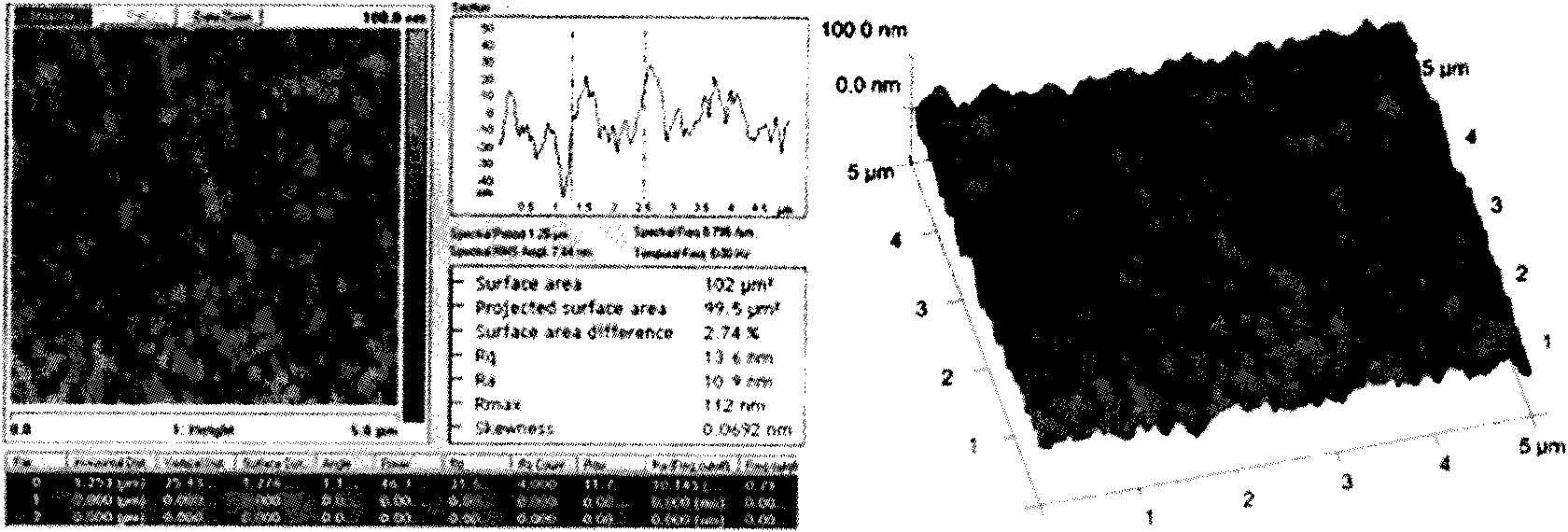
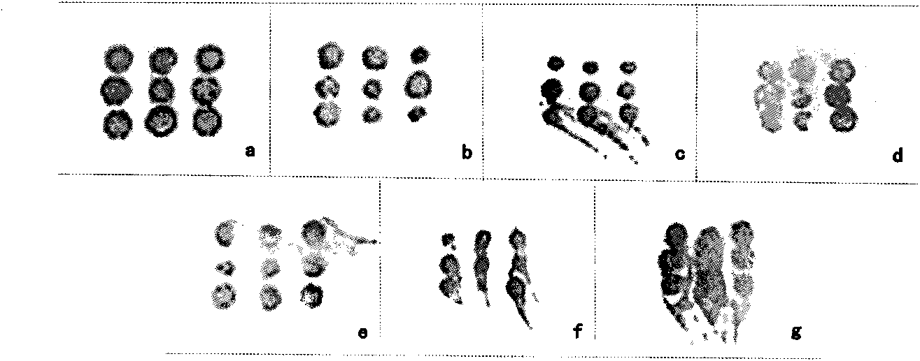
[0071] Preparation of slides with seven different modification methods and comparison of fixation effects.
[0072] 1. Commercially available glass slides were washed with strong alkali and concentrated sulfuric acid, soaked in ultrapure water overnight, and modified by the following methods.
[0073](1) Preparation of APES-modified glass slides: APES is ready-to-use and ready-to-use. Put the cleaned slides into APES diluted with 1:50 acetone, stay for 20-30s, take it out and pause for a while, then put it into pure acetone solution to remove unbound APES, and immediately immerse in pure water for 2-5 minutes Rinse, store in a dust-free place away from light, and dry in an oven at 80°C on a slide rack.
[0074] (2) Preparation of poly-lysine-modified glass slides: put the cleaned and dried glass slides into poly-lysine solution diluted with deionized water at a ratio of 1:10, shake on a shaker, double Rinse with steamed water, soak for 5 minutes, bake in an oven at 60°C for ...
Embodiment 3
[0084] Antibody microarrays were prepared using the above-mentioned agarose-modified slide glass as a substrate.
[0085] 1. Chip structure design
[0086] The chip structure is as Figure 4 As shown, it includes the chip carrier (1), the agarose gel layer (2) covered on the chip carrier (1), and the agarose gel layer (2) is fixed with two rows and four columns, a total of eight 4×4 Antibody microarray (3), the sample volume is 50-70 nl, and the spot diameter is 500-600 μm. Each microarray is separated by a chip-specific fence or a Super PAP Pen scribe line (4). The chip carrier is a standard glass slide, and a label area (5) can be reserved at one end of the glass slide.
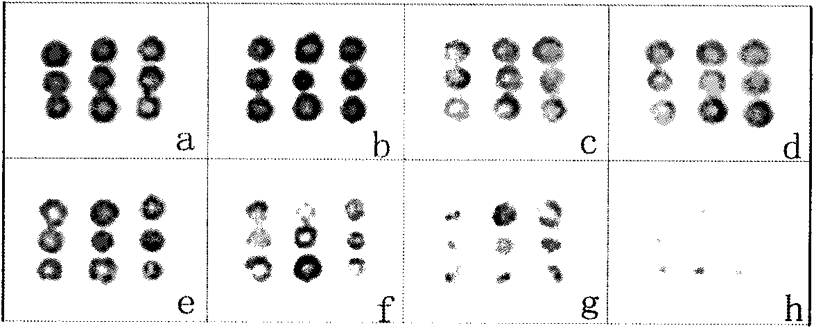
[0087] 2. Selection of the optimal concentration of capture antibody
[0088] (1) Dilute the capture antibody (rabbit anti-LCDV antibody or rabbit anti-WSSV antibody) with PBS buffer solution containing 50% glycerol at pH 7.4, so that the concentration is 1, 0.5, 0.1, 0.05, 0.01, 0.005, 0.001, 0.0005 m...
PUM
 Abstract
Description
Claims
Application Information
 - R&D
- Intellectual Property
- Life Sciences
- Materials
- Tech Scout
- Unparalleled Data Quality
- Higher Quality Content
- 60% Fewer Hallucinations
Browse by: Latest US Patents, China's latest patents, Technical Efficacy Thesaurus, Application Domain, Technology Topic, Popular Technical Reports.
© 2025 PatSnap. All rights reserved.Legal|Privacy policy|Modern Slavery Act Transparency Statement|Sitemap|About US| Contact US: help@patsnap.com


