Polyethylene glycol modified human serum albumin and preparation method thereof
A human albumin and polyethylene glycol technology, applied in the preparation method of peptides, serum albumin, albumin peptides, etc., can solve problems such as prolonging the dosing cycle, reduce the frequency of dosing, prolong the half-life, and reduce the source of remission nervous effect
- Summary
- Abstract
- Description
- Claims
- Application Information
AI Technical Summary
Problems solved by technology
Method used
Image
Examples
Embodiment 1
[0039] Site-directed Single Modification of Human Serum Albumin by Modifier mPEG-MAL
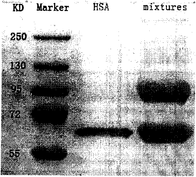
[0040]mPEG-MAL (Mr=20kD) and human serum albumin (based on protein content) were dissolved in pH 6.5, 50mmol / L phosphate buffer (containing 10mmol / L EDTA) at a molar ratio of 2:1, shaken in a water bath at 30°C Bed, shake gently for 20h. Take 3 μL of the modified reaction solution for SDS-PAGE detection, see the staining results figure 1 . figure 1 It shows that compared with HSA, a new band appears in the modified reaction solution lane (mixtures lane), and the molecular weight is about 90kD. The molecular weight of the PEG modifier used in the modification reaction is 20kD, and the molecular weight of albumin is 66.5kD, so it can be inferred that the newly appeared band is PEG-cys 34 -HSA, and one HSA molecule is connected with one PEG chain.
Embodiment 2
[0042] mPEG site-directed single-modification product of cysteine 34 of albumin (PEG-cys 34 - Separation and purification of HSA)
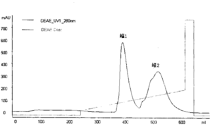
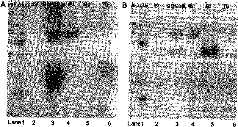
[0043] The modified product was separated by DEAE Sepharose FF chromatography medium. The modified reaction solution was diluted 5 times with pH 6.5, 10mmol / L phosphate buffer solution, and then loaded. The flow rate is 5.0ml / min, and the detection wavelength is 280nm. The elution curve shows two elution peaks, which are collected separately, desalted and concentrated by ultrafiltration, and the purity of the sample is detected by SDS-PAGE electrophoresis. The electrophoresis film is stained with iodine first and then stained with iodine. Both PEG and protein-bound PEG can be stained; Coomassie brilliant blue staining can stain proteins, and the bands that can be stained by both dyes are PEG-cys 34 -HSA. DEAE chromatograms and SDS-PAGE spectra are shown in figure 2 and image 3 . image 3 Display: DEAE peak 1 (swimming lane 4) in the iod...
Embodiment 3
[0046] PEG-cys 34 -HSA and HSA secondary structure determination
[0047] Determination of PEG-cys by Circular Dichroism (CD) 34 -HSA and the secondary structure of HSA. Weigh 10mgHSA and 9mg PEG-cys respectively 34 -HSA, dissolved in pH 7.4, 50mmol / L phosphate buffer, so that the final protein concentration is 5μmol / L. Use a 1mm colorimetric cell to measure HSA and PEG-cys at 25°C 34 - CD spectrum of HSA in the wavelength range 190-260 nm. The secondary structure of HSA and PEG-cys34-HSA was calculated by random special software. HSA and PEG-cys 34 Circular Dichroism (CD) of -HSA see Figure 4 . Depend on Figure 4 It can be seen that HSA and PEG-cys 34 - The CD spectrum of HSA has a similar peak shape, and the CD spectrum of the two is almost completely overlapped, which shows that the secondary structure of the two is almost completely consistent, and the secondary structure calculated by the random software has further confirmed this (see Table 1 ), suggesting t...
PUM
| Property | Measurement | Unit |
|---|---|---|
| Molecular weight | aaaaa | aaaaa |
| Molecular weight | aaaaa | aaaaa |
| Molecular weight | aaaaa | aaaaa |
Abstract
Description
Claims
Application Information
 - R&D
- Intellectual Property
- Life Sciences
- Materials
- Tech Scout
- Unparalleled Data Quality
- Higher Quality Content
- 60% Fewer Hallucinations
Browse by: Latest US Patents, China's latest patents, Technical Efficacy Thesaurus, Application Domain, Technology Topic, Popular Technical Reports.
© 2025 PatSnap. All rights reserved.Legal|Privacy policy|Modern Slavery Act Transparency Statement|Sitemap|About US| Contact US: help@patsnap.com


