
Fluorescence navigation system used in tumor surgery

A navigation system and fluorescence technology, applied in the field of clinical medical surgery, can solve problems such as the difficulty of identifying tumor metastasis and lymph node tracking in residual tumor tissue, and achieve the effect of improving the effect of surgical treatment and reducing the mortality rate.

Inactive Publication Date: 2011-11-09
SHANXI MEDICAL UNIV
View PDF0 Cites 2 Cited by
  • Summary
  • Abstract
  • Description
  • Claims
  • Application Information

AI Technical Summary

Problems solved by technology

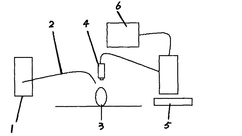
[0004] In order to solve the problems of tumor boundary identification, residual tumor tissue identification and tumor metastatic lymph node tracking during the existing surgical tumor resection, the present invention provides an intraoperative fluorescence navigation system for tumor surgery and realizes a new surgical technique. Medium Imaging Mode

Method used

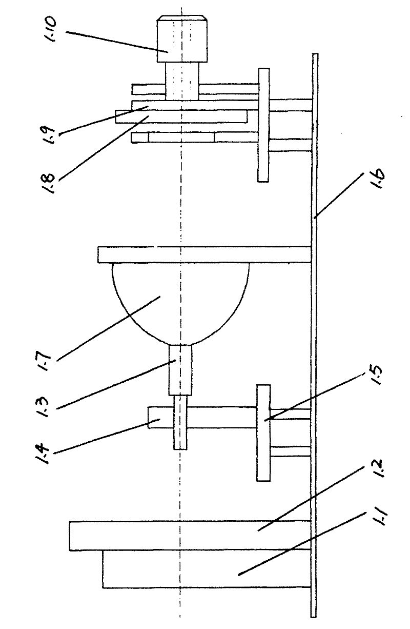

Image

  • Fluorescence navigation system used in tumor surgery
  • Fluorescence navigation system used in tumor surgery
  • Fluorescence navigation system used in tumor surgery

Examples


Embodiment 1

[0057] Using liposome 2000-mediated gene transfection, the chickenβ-actin-DsRed-Neo vector was transfected into the mouse bladder cancer BTT739 cell line; G418 screening was used to obtain a monoclonal cell line stably expressing red fluorescent protein (DsRed) (BTT739-DsRed); establishment of xenograft tumor model; fluorescence navigation during tumor surgery to record fluorescence images and fluorescence intensity related to tumor growth and metastasis. The results showed that the DsRed-labeled BTT-739 cell line was red fluorescence under the fluorescence microscope, and the cells were planted subcutaneously in mice. On the 7th day after the establishment of the mouse bladder cancer xenograft model, the fluorescence navigation during tumor surgery, the excitation light wavelength 560-580nm, the emission wavelength was 590-610nm, the tumor showed red fluorescence, and the fluorescence intensity of the tumor gradually increased. Local lymph node metastasis was observed on the 2...

Embodiment 2

[0060] Fluorescent substances have a good affinity with tumor tissues. After intravenous injection or oral administration of photosensitizers (48-72 hours), patients are then exposed to light, and the fluorescence spectral characteristic curve can be recorded to determine the location of the tumor. In addition, autofluorescence spectrum diagnosis does not require additional photosensitizing agents, and uses the fluorescence generated by human tissues under laser light to perform spectral analysis to distinguish tumors. It is a non-invasive and rapid diagnostic technique without oral or injection of photosensitizers.

Embodiment 3

[0062] In fluorescent molecular imaging, in order to identify biomolecular processes of interest, it is necessary to use fluorescent molecular probes for observation and quantitative analysis. In addition, there are fluoresceins labeled with antibodies, and fluorescein isothiocyanate is a protein-binding , the maximum absorption wavelength is 490-495nm, the maximum emission wavelength is 520-530nm, showing bright yellow-green fluorescence; tetramethyl rhodamine isothiocyanate, the maximum absorption wavelength is 550nm, and the maximum emission wavelength is 620nm, Shows orange-red fluorescence.


PUM


Abstract

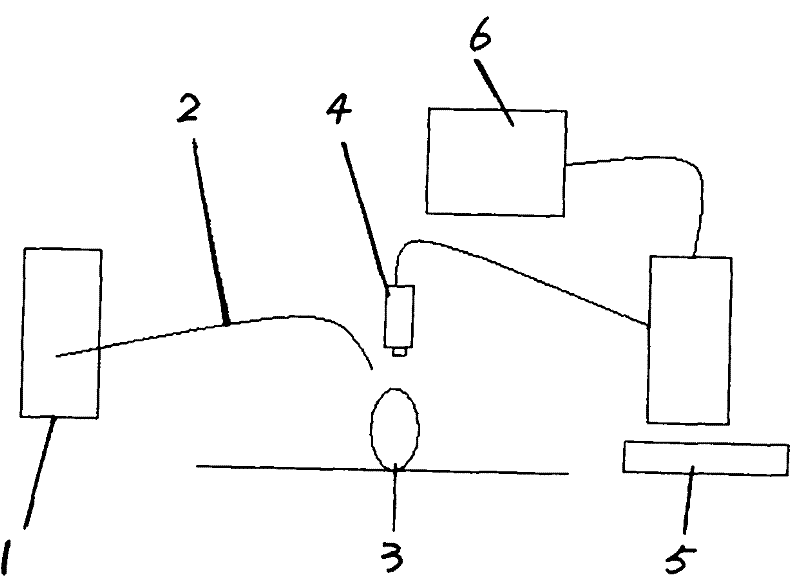
The present invention relates to a fluorescence navigation system used in tumor surgery, which belongs to clinical medicine surgery. The present invention solves problems existing in the surgical excision operation process of tumors that tumor boundaries are difficult to identify, remnant tumor tissue is difficult to recognize, and tumor metastasis lymph glands are difficult to track. The fluorescence navigation system used in tumor surgery is characterized in that the system comprises a biofluorescence excitation light source, a light guide system, a specificity targeted marking fluorescent material, a video camera and an image display and processing device connected with the video camera. The present invention provides fluorescence navigation concept for tumor surgery based on the priorart. When physicians identify tumor boundaries, recognize remnant tumor tissue, and track tumor metastasis lymph glands with a fluorescence observing mirror or a camera display system, tumor tissue is thoroughly excited, operation therapeutic effect is effectively improved, and the death rate of tumor patients is reduced. The present invention makes a breakthrough in tumor surgery.

Description

technical field [0001] The invention belongs to the field of clinical medicine and surgery, in particular to a fluorescence navigation system in tumor surgery. Background technique [0002] According to the World Health Organization, 7,600,000 people died of cancer in 2005, accounting for 13% of all 58,000,000 deaths. For example, there are 1.3 million non-skin cancer patients in the United States every year, half of which are cured by surgical resection, about 5% are cured by chemotherapy and / or radiotherapy, and the remaining patients cannot be cured. The main reason why it cannot be cured is that the tumor cannot be completely removed by surgery, for example, the tumor range cannot be correctly identified during the operation, and the micrometastases cannot be clearly identified. [0003] However, the existing intraoperative imaging examinations limit its application due to various factors. For example, lipiodol angiography can see the blood vessel structure clearly, bu...

Claims


Application Information

Patent Timeline
Patent Type & Authority Patents(China)
IPC IPC(8): G03B15/03G03B15/14
Inventor 杨晓峰
Owner SHANXI MEDICAL UNIV
Learn More