
Nano magnetic resonance imaging contrast agent and preparation method thereof

A technology of magnetic resonance imaging and contrast agent, which is applied in the direction of MRI/magnetic resonance imaging contrast agent, etc.

Inactive Publication Date: 2010-09-15
HUNAN UNIV
View PDF8 Cites 8 Cited by
  • Summary
  • Abstract
  • Description
  • Claims
  • Application Information

AI Technical Summary

Problems solved by technology

Macromolecules used to coat magnetic particles have been reported to include proteins US4675173A1, US4849210A, CN1724076A, sugars US5766572A, US2007116955A1, antibodies WO2008063371A2, amino acids US6207134B1, polyvinylpyrrolidone US2006204438A1, etc., but no magnetic substances modified with humic acids have been seen. Reports on Particles Used as Contrast Agents

Method used


Image

  • Nano magnetic resonance imaging contrast agent and preparation method thereof
  • Nano magnetic resonance imaging contrast agent and preparation method thereof
  • Nano magnetic resonance imaging contrast agent and preparation method thereof

Examples


Embodiment 1

[0034] Example 1: In a 100mL three-necked flask with mechanical stirring, reflux condenser, temperature controller and argon protection, add 50mL ultrapure water, 0.3g sodium humate, 10mL concentrated ammonia, and heat to 70℃, Immediately add 5 mL of an ultrapure aqueous solution containing 4mmoL of ferric sulfate and 2mmoL of manganese acetate, heat at 70°C for 2 hours, and then cool to room temperature. The product is centrifuged, washed with ultrapure water for 3 to 5 times, and then subjected to semipermeable membrane dialysis. After separation and purification, it was dispersed in physiological saline to prepare 0.005M humic acid-coated manganese ferrite magnetic resonance imaging contrast agent.

Embodiment 2

[0035] Example 2: In a 100mL three-necked flask with mechanical stirring, reflux condenser, temperature controller and argon protection, add 50mL ultrapure water, 0.3g sodium fulvic acid, 10mL concentrated ammonia, and heat to 70℃ immediately Add 5 mL of an ultrapure aqueous solution containing 4mmoL of ferric citrate and 2mmoL of cobalt nitrate, keep it at 80°C for 2 hours and then cool to room temperature. The product is centrifuged, washed with ultrapure water 3 to 5 times, and then separated by a silica gel column. After purification, it was dispersed in ultrapure water to prepare a 0.05M fulvic acid-coated cobalt ferrite magnetic resonance imaging contrast agent.

Embodiment 3

[0036] Example 3: In a 500mL three-necked flask with mechanical stirring, reflux condenser, temperature controller and argon protection, add 250mL ultrapure water, 2g humic acid, 2g sodium hydroxide, and raise the temperature to 100℃ immediately Add 25mL of an ultrapure aqueous solution containing 20mmoL of ferric chloride and 10mmoL of ferrous chloride, reflux for 2 hours at 100°C, and then cool to room temperature. The product is centrifuged, washed with ultrapure water 3 to 5 times, and then passed through a silica gel layer. After column separation and purification, it was dispersed in glucose injection to prepare 0.05M humic acid-coated ferroferric oxide magnetic resonance imaging contrast agent.


PUM

PropertyMeasurementUnit
particle diameteraaaaaaaaaa
diameteraaaaaaaaaa

Abstract

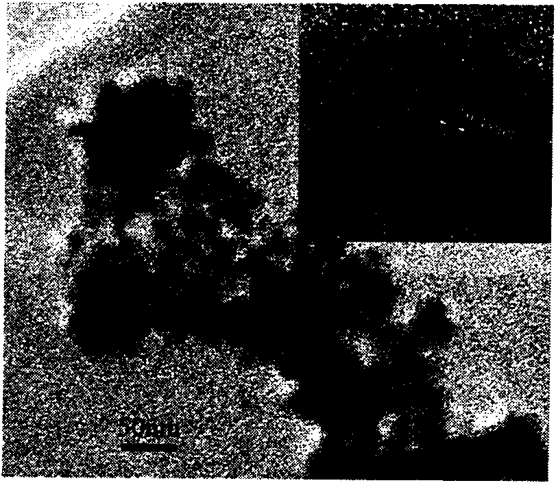
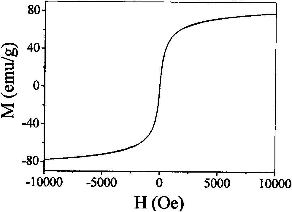
The invention relates to a nano magnetic resonance imaging contrast agent and a preparation method thereof. The contrast agent consists of external aqueous phase and humic acid substance-modified super-paramagnetic ferrite nano crystal dispersed in the external aqueous phase. The super-paramagnetic ferrite nano crystal is synthesized by adopting a chemical co-precipitation method, and the grain diameter is 15 to 30 nanometers. The nano crystal is purified and then dispersed in the external aqueous phase to form the contrast agent for intravenous injection. The contrast agent can be stabilized for over two years. The contrast agent has low toxicity, good biocompatibility, large time window and high saturation magnetization, and the half-life period of the contrast agent in the blood reaches 30 to 60 minutes. The contrast agent is absorbed by netlike endothelial systems in liver and spleen tissues through intravenous injection, and is focused in the liver and spleen tissues so as to shorten the T2 relaxation time. In a T2 image, signals of normal liver and spleen tissues are obviously reduced, and signals of tumor or pathological change tissues have no obvious change, so the contrast agent improves the imaging contrast of the focus and the normal tissue and can be used for early diagnosis of pathological changes such as micro tumor of liver and spleen tissues and the like.

Description

Technical field [0001] The invention relates to a nano magnetic resonance imaging contrast agent, in particular to a liver and spleen nano magnetic resonance imaging contrast agent and a preparation method thereof. Background technique [0002] Magnetic resonance imaging (MRI) technology has now developed into a common method in clinical medical diagnosis and is one of the most effective methods for diagnosing tumors. Because it is non-invasive and the patient is not exposed to potentially harmful radiation, this technology is becoming more and more popular. In order to enhance the contrast and clarity between the image of the diseased tissue and the normal tissue, it is necessary to select a suitable contrast agent to display anatomical features. Magnetic resonance imaging contrast agent is a medical diagnostic reagent that can be injected intravenously into the human body to shorten imaging time, improve imaging contrast and clarity, and display the functional status of tissue...

Claims


Application Information

Patent Timeline
Patent Type & Authority Applications(China)
IPC IPC(8): A61K49/06
Inventor 邹炳锁周春姣容鹏飞王维万强周建大张声旺张文杰
Owner HUNAN UNIV
Learn More