Method for preparing dried amnion
An amniotic membrane drying technology, applied in medical science, prostheses, etc., can solve the problems that the effect has not been confirmed, and achieve the effect of complete cell phenotype and basement membrane, easy implementation, and optimized amniotic membrane characteristics
- Summary
- Abstract
- Description
- Claims
- Application Information
AI Technical Summary
Problems solved by technology
Method used
Image
Examples
Embodiment 1
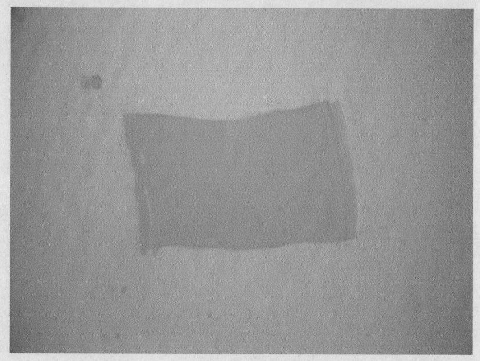
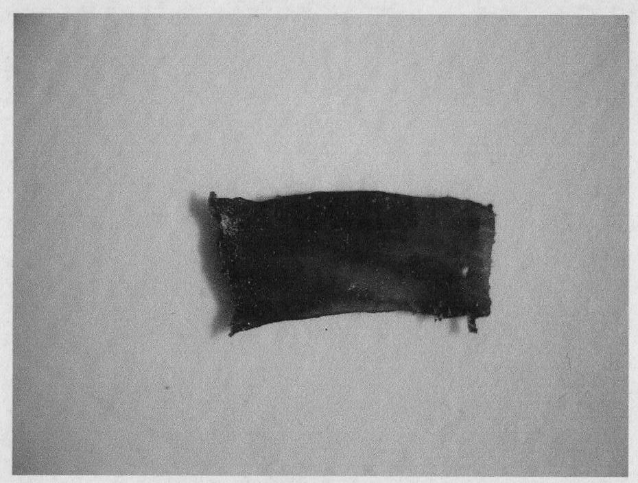
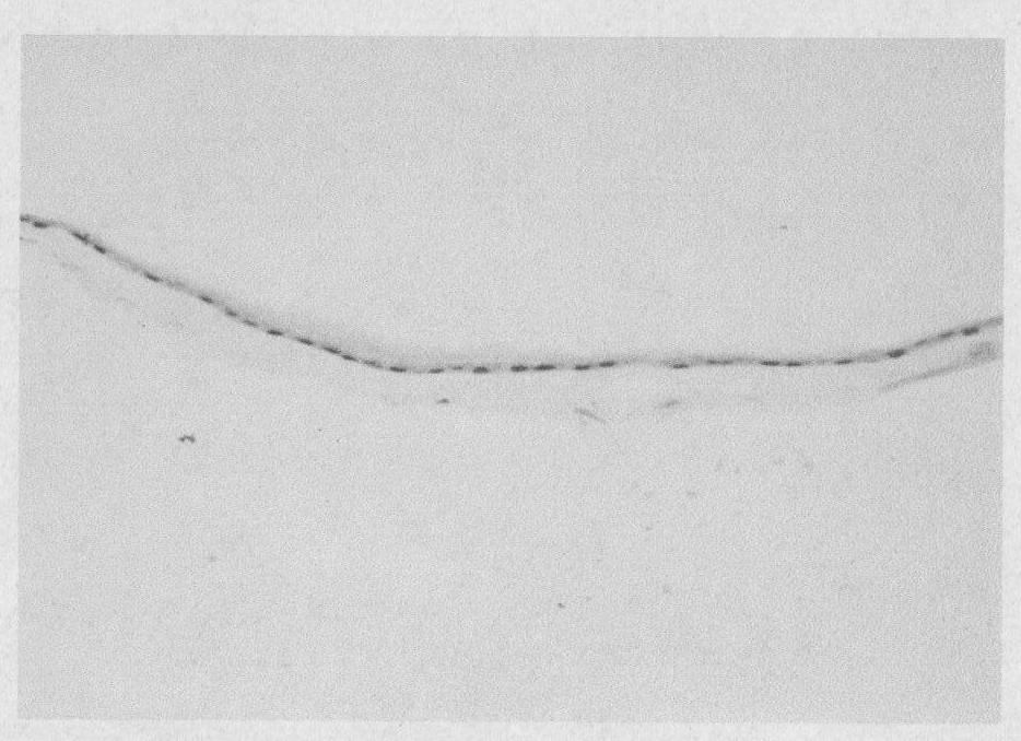
[0038] The preparation method of the present invention comprises the following steps: the donor is taken from the fetal membrane obtained by cesarean section, and the preoperative virological test results of the puerpera are all negative; Bacterial saline was used to wash the blood on the surface; the operator wore sterile gloves on both hands, gently separated the amniotic membrane from the chorionic membrane, rinsed it with sterile BSS solution, spread it on a sterile glass plate, and scraped it with a cell scraper. Remove the spongy layer, fibroblast layer and serous exudate; soak and disinfect the fresh amniotic membrane with PBS containing 50,000 U / L penicillin, 80,000 U / L tobramycin, and 100 mg / L streptomycin for 3 Time × 5min; at -20°C-4°C, soak the sterile amniotic membrane in ECM glue for 15min-30min; drop 0.25% sterile sodium fluorescein solution 0.2ml in the fume hood at 16°C-37°C Stain the amniotic membrane for 5 minutes, then lay the dyed amniotic membrane on the ...
Embodiment 2
[0040] The preparation method of the present invention comprises the following steps: the donor is taken from the fetal membrane obtained by cesarean section, and the preoperative virological test results of the puerpera are all negative; Bacterial saline was used to wash the blood on the surface; the operator wore sterile gloves on both hands, gently separated the amniotic membrane from the chorionic membrane, rinsed it with sterile BSS solution, spread it on a sterile glass plate, and scraped it with a cell scraper. Remove the spongy layer, fibroblast layer and serous exudate; soak and disinfect the fresh amniotic membrane with PBS containing 50,000 U / L penicillin, 80,000 U / L tobramycin, and 100 mg / L streptomycin for 3 Time × 5min; at -20°C-4°C, soak the sterile amniotic membrane in FN glue for 15min-30min; drop 0.1% sterile trypan blue solution 0.2ml in the fume hood at 16°C-37°C Stain the amniotic membrane for 5 minutes, then lay the dyed amniotic membrane on the nitrocell...
Embodiment 3
[0042] The preparation method of the present invention comprises the following steps: the donor is taken from the fetal membrane obtained by cesarean section, and the preoperative virological test results of the puerpera are all negative; Bacterial saline was used to wash the blood on the surface; the operator wore sterile gloves on both hands, gently separated the amniotic membrane from the chorionic membrane, rinsed it with sterile BSS solution, spread it on a sterile glass plate, and scraped it with a cell scraper. In addition to its spongy layer, fibroblast layer and serous exudate. Soak fresh amnion in PBS containing 50,000 U / L penicillin, 80,000 U / L tobramycin, and 100 mg / L streptomycin for 3 times×5min; Soak the amniotic membrane in Matrige gel for 15min-30min; in a fume hood at 16°C-37°C, drop 0.2ml of 0.5% sterile tiger red solution on the amnion and stain it for 5min, then spread the stained amnion on the nitrocellulose membrane , in a sterile fume hood to dry natur...
PUM
| Property | Measurement | Unit |
|---|---|---|
| thickness | aaaaa | aaaaa |
| thickness | aaaaa | aaaaa |
| thickness | aaaaa | aaaaa |
Abstract
Description
Claims
Application Information
 - R&D
- Intellectual Property
- Life Sciences
- Materials
- Tech Scout
- Unparalleled Data Quality
- Higher Quality Content
- 60% Fewer Hallucinations
Browse by: Latest US Patents, China's latest patents, Technical Efficacy Thesaurus, Application Domain, Technology Topic, Popular Technical Reports.
© 2025 PatSnap. All rights reserved.Legal|Privacy policy|Modern Slavery Act Transparency Statement|Sitemap|About US| Contact US: help@patsnap.com


