

Spherical-cavity microelectrode array biosensor and preparation method thereof
A microelectrode array and biosensor technology is applied in the field of preparation of spherical cavity microelectrode array biosensors to achieve the effects of improving signal-to-noise ratio, high sensitivity and increasing loading capacity
- Summary
- Abstract
- Description
- Claims
- Application Information
AI Technical Summary
Problems solved by technology
Method used
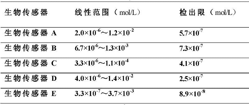
Image

Examples
Embodiment 1
[0034] For example, the platinum electrode was ultrasonically washed with acetone, ethanol, and water for 30 minutes and then stored in water. A polystyrene microsphere emulsion with a mass fraction of 1% was prepared with a 1:0.5 ethanol:water mixture as a solvent, and the size of the microspheres was 1500 nm. Using a mixture of water and ethanol with a volume ratio of 8.5:0.85 as the subphase, the polystyrene microsphere emulsion was slowly dripped onto the surface of the subphase. After standing for 15 minutes, the sliding barrier was compressed at a speed of 10 cm / min. When the membrane pressure reaches 10mN / m, keep the same membrane pressure for 30 minutes to stabilize the Langmuir membrane on the gas-liquid interface. The polystyrene microsphere array electrode was obtained by using the vertical pulling method to transfer it to the electrode at a speed of 1mm / min.
[0035] Mix 5mL of ethyl orthosilicate with 60mL of ethanol, put it in a constant temperature magnetic stirre...
Embodiment 2
[0038] The indium tin oxide electrode was ultrasonically washed with acetone, ethanol, and water for 30 minutes and then stored in water. A polystyrene microsphere emulsion with a mass fraction of 1% was prepared with a 1:1 ethanol:water mixture as a solvent, and the size of the microspheres was 1500 nm. Using a mixture of water and ethanol with a volume ratio of 8.5:1 as the subphase, the polystyrene microsphere emulsion was slowly dripped onto the surface of the subphase, and after standing for 20 minutes, the sliding barrier was compressed at a speed of 20 cm / min. When the membrane pressure reaches 14mN / m, keep the same membrane pressure for 30 minutes to stabilize the Langmuir membrane on the gas-liquid interface. The two-dimensional polystyrene microsphere array electrode is obtained by using the vertical pulling method to transfer it to the electrode at a speed of 1mm / min.
[0039] Prepare a mixture of 0.3mol / L zinc nitrate and 0.1mol / L citric acid, and heat the mixture to...
Embodiment 3
[0042] The glassy carbon electrode was ultrasonically washed with acetone, ethanol, and water for 30 minutes and then stored in water. A polystyrene microsphere emulsion with a mass fraction of 1% was prepared using a mixture of ethanol and water with a volume ratio of 1:1.5 as a solvent, and the size of the microspheres was 1500 nm. Using a mixture of water and ethanol with a volume ratio of 8.5:1.5 as the subphase, the polystyrene microsphere emulsion was slowly dripped onto the surface of the subphase, and after standing for 25 minutes, the sliding barrier was compressed at a speed of 30 cm / min. When the membrane pressure reaches 18mN / m, keep the same membrane pressure for 30 minutes to stabilize the Langmuir membrane on the gas-liquid interface. The two-dimensional polystyrene microsphere array electrode is obtained by using the vertical pulling method to transfer it to the electrode at a speed of 1mm / min.
[0043] Take 20mL of tetra-n-butyl titanate and add dropwise to 80mL...
PUM
| Property | Measurement | Unit |
|---|---|---|
| Size | aaaaa | aaaaa |
Abstract
Description
Claims
Application Information