Preparation method of membrane protein polypeptides for improving detection rate of membrane proteins

A technology of membrane protein and detection rate, applied in the field of preparation in the field of biological detection technology, achieves the effects of low cost, short time consumption and simplified preparation steps

Inactive Publication Date: 2012-11-28
SHANGHAI JIAOTONG UNIV
View PDF0 Cites 0 Cited by
  • Summary
  • Abstract
  • Description
  • Claims
  • Application Information

AI Technical Summary

Problems solved by technology

[0005] The purpose of the present invention is to overcome the technical difficulties in the pretreatment of complex cell membrane protein spectrum analysis, and to provide a method for preparing membrane protein polypeptides that improves the detection rate of membrane proteins

Method used


Image

  • Preparation method of membrane protein polypeptides for improving detection rate of membrane proteins
  • Preparation method of membrane protein polypeptides for improving detection rate of membrane proteins
  • Preparation method of membrane protein polypeptides for improving detection rate of membrane proteins

Examples


Embodiment 1

[0023] Kunming rats (male, weighing 250-300 grams, purchased from Shanghai Slack Experimental Animal Center) took blood from the orbit, put it into a test tube previously filled with 2% potassium oxalate solution, and mixed quickly. Centrifuge at 1000g at 4°C for 5 minutes, and use a pipette to remove the supernatant and the white blood cell film on the upper layer of red blood cells. Slowly mix the precipitated erythrocytes with twice the volume of normal saline, centrifuge again at 1000 g at 4°C for 5 min, discard the supernatant, add normal saline and mix well, and repeat centrifugation for 4 to 5 times. After the last centrifugation of the red blood cells, discard the supernatant, which is the washed red blood cells. Draw 50 μl of red blood cells, add 2450 μl of pre-cooled 1% glutaraldehyde solution (the final concentration of red blood cells is 2%) in an ice bath, and slowly rotate and react at 4°C for 0.5 hours. After the reaction, centrifuge at 1000 g for 5 min at 4°C....

Embodiment 2

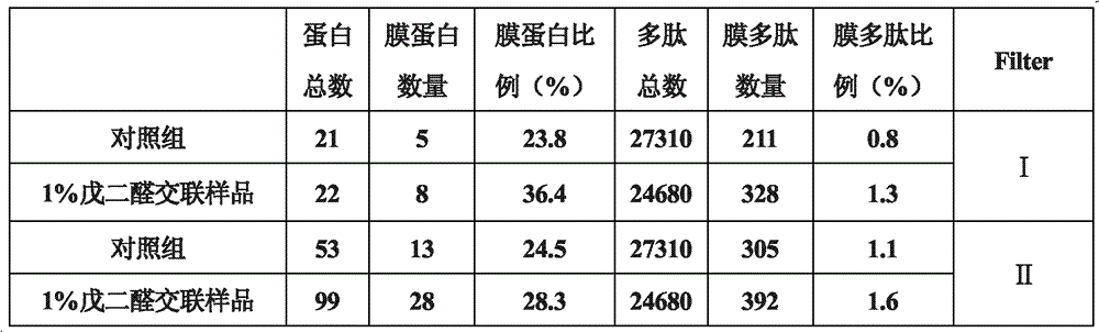
[0027] Take 50 μl of washed mouse red blood cells, add 2450 μl of pre-cooled 0.1% glutaraldehyde solution (final concentration of red blood cells is 2%) in an ice bath, and slowly rotate and react at 37° C. for 0.5 hours. After the reaction, centrifuge at 1000 g for 5 min at 4°C. The precipitated cells were washed 3 times with normal saline in the same way. Erythrosomes cross-linked with 0.1% glutaraldehyde were obtained. Take 10 μl each of untreated erythrocytes, 1% glutaraldehyde cross-linked and solidified erythrocytes prepared in Example 1, and 0.1% glutaraldehyde cross-linked erythrocytes (about 8.7×10 7 ), add 490 μl 10 mmol PBS (pH7.4), add 10 μl 0.1 μg / μl sequencing grade trypsin. Enzymatic hydrolysis in a 37°C oven for 14-16 hours. Collect the enzymatic peptides, use C18Zip-tips to desalt and enrich the peptides. Samples were lyophilized for LC-LTQ analysis. The obtained mass spectrometry data was searched using SEQUEST; Trans-Proteomie Pipeline (ISB / SPC Proteomi...

Embodiment 3

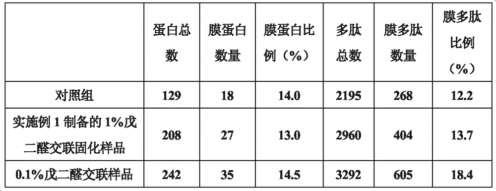
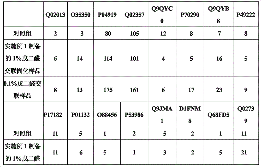
[0036]Get each 10 μl of untreated erythrocytes and 0.1% glutaraldehyde cross-linked and solidified erythrocytes prepared in Example 2 (about 8.7×10 7 ), add 490 μl 50 mmol Tris-HCl buffer, pH 8.0, add 10 μl 0.1 μg / μl sequencing grade endoproteinase GluC (Endoproteinase Glu-C). Enzymatic hydrolysis at 25°C for 14-16 hours. Collect the enzymatic peptides, use C18 Zip-tips to desalt and enrich the peptides. Samples were lyophilized for LC-LTQ analysis. The obtained mass spectrum data was searched using SEQUEST; Filter was set to ΔCn>=0.100; Xcorr(±1, 2, 3)=1.90, 2.20, 3.75; #matches=1. Results A higher proportion of membrane proteins was detected in the sample prepared in Example 2 and treated with 0.1% glutaraldehyde cross-linking and curing. Table 5 shows the number and ratio of membrane polypeptides and membrane proteins detected before and after glutaraldehyde crosslinking:

[0037] table 5

[0038]


PUM


Abstract

The invention relates to a preparation method of membrane protein polypeptides for improving the detection rate of membrane proteins in the technical field of a biological detecting. The method comprises the following steps: crosslinking and solidifying cytoplasm proteins: firstly, processing integral cells by utilizing a glutaraldehyde solution, enabling the cytoplasm proteins to be mutually crosslinked into a dense solid; centrifugally collecting glutaraldehyde crosslinking and solidification cells; suspending the glutaraldehyde crosslinking and solidification cells by utilizing a proteolysis reaction solution; adding proteases to carry out an enzymolysis reaction; collecting enzymolysis polypeptides; carrying out the desalting and the enriching; preparing the desalted enzymolysis polypeptides; directly carrying out the LC-MS (liquid chromatogram-mass spectrometer) / MS detection by utilizing the enzymolysis polypeptides; and analyzing the obtained mass spectrum data to obtain the needed membrane protein information. The method provided by the invention can rapidly obtain the more and more reliable membrane protein information and effectively reduces the signal interference of thecytoplasm proteins in the LC-MS / MS detection.

Description

technical field [0001] The invention relates to a preparation method in the technical field of biological detection, in particular to a preparation method of a membrane protein polypeptide which improves the detection rate of the membrane protein. Background technique [0002] The cell membrane is the basis of cell shape and the barrier between cells and the external environment. Membrane proteins are bridges for the transmission of substances, energy and information inside and outside cells, and undertake important biological functions. Therefore, the analysis of cell membrane proteins is the basis for studying scientific issues such as health and disease, environmental changes, and genetic abnormalities. As a relatively new technology, membrane proteomics has been widely used in the research of revealing cell function, explaining basic biological problems and discovering new drug targets. In particular, the use of analytical methods such as mass spectrometry and liquid c...

Claims


Application Information

Patent Timeline
Patent Type & Authority Patents(China)
IPC IPC(8): G01N30/08C12P21/06
Inventor 李荣秀徐泓马贵军邹琦程超
Owner SHANGHAI JIAOTONG UNIV
Why Eureka
Try Eureka