Method and detector for detecting tumor microsomes by using laser tweezers and micro fluidics

A technology of laser tweezers and microsomes, which is applied in the direction of instruments, measuring devices, scientific instruments, etc., can solve the problems of insufficient sensitivity and inapplicability for early diagnosis of tumors, and achieve the effect of broad market prospects, easy operation, and rapid detection

Inactive Publication Date: 2011-11-02
嘉兴文斌生物技术有限公司 +1
View PDF6 Cites 9 Cited by
  • Summary
  • Abstract
  • Description
  • Claims
  • Application Information

AI Technical Summary

Problems solved by technology

Biochemical methods such as ELISA are not suitable for early diagnosis of tumors due to their instability and insufficient sensitivity

Method used


Image

  • Method and detector for detecting tumor microsomes by using laser tweezers and micro fluidics
  • Method and detector for detecting tumor microsomes by using laser tweezers and micro fluidics

Examples


preparation example Construction

[0039] Wherein, the preparation method of antibody particles bound to tumor microsomes comprises the following steps:

[0040] Step f: biotin-label the pre-purchased tumor microsomal antibody with EZ-Link NHS-PEG12-Biotin kit;

[0041] Step g: Linking biotin-labeled tumor microsomal antibody to streptavidin polymer particles to form tumor microsomal antibody-bound particles.

[0042] The preparation method of the microfluidic chip comprises the following steps:

[0043] Step i: Draw the mask layout of the microfluidic chip by drawing software, print it with a laser printer with a resolution of 12000dpi, and make a photolithographic mask plate on the transparent film.

[0044] Step j: placing the glass substrate in acetone and alcohol for ultrasonic treatment, then boiling in concentrated H2SO4, rinsing with deionized water, drying with nitrogen, and drying with heat to remove water vapor;

[0045] Step k: Spin-coat photoresist on the surface of the cleaned glass substrate, s...

Embodiment 2

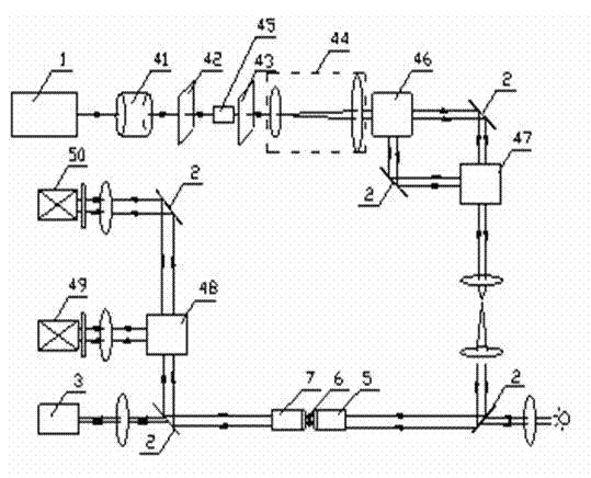
[0051] Laser tweezers and microfluidic detectors for tumor microsomes, such as figure 1 As shown, it includes laser system 1, nanopositioning system 2, camera system 3, optical components 4, mechanical components, data acquisition and transmission system, automatic control and image processing application software. Laser system 1 is a diode-pumped solid-state laser transmitter or fiber laser transmitter (power 1 to 5 watts), camera system 3 is an IXON electron multiplier charge coupler, nanopositioning system 2 is a nanoscale steerable mirror, optical components 4 It includes a Faraday isolator 41, half-wave plates 42, 43, lenses 44, polarizing beam splitters 45, 46, 47, 48, position-sensitive photodetectors 49, 50, microfluidic chip tube cavity 6, mechanical components including shutters, Eyepiece console and automatic sample console.

[0052] A semiconductor pumped laser (diode pumped solid state, DPSS, 1064 nm, 4W, continuous wave mode laser) was used as the trapping laser...


PUM


Abstract

The invention relates to a method and equipment for detecting tumor microsomes, and especially relates to and discloses a method and a detector for detecting tumor microsomes by using laser tweezers and micro fluidics. The detection method comprises the steps that: laser tweezers of different polarizations are generated; dual-beam double laser tweezers are used for focusing, and particles combined with tumor microsome antibodies in a channel of a micro fluidic chip are captured by the tweezers; a sample to be detected is mixed with the particles combined with tumour microsome antibodies; changes of physical parameters of the particles combined with tumor microsome antibodies before and after being mixed with the sample are collected by the detector, and qualification and accurate quantitation of the sample requiring detection can be carried out according to the parameters. The detector comprises a laser system, a nanometer positioning system, a camera system, optical parts, mechanical parts, a data collecting and transmitting system, and automatic controlling and image processing application software. According to the invention, single tumor microsome particle can be detected, and the method and the equipment are characterized in high specificity, simple operation, short detection period, high sensitivity, and good repeatability. Also, with the method and the equipment, blood samples can be re-detected.

Description

technical field [0001] The invention relates to a detection method and detection equipment for tumor microsomes, in particular to a laser tweezers and microfluidic detection method for tumor microsomes and a detector. Background technique [0002] In recent decades, the incidence of cancer has been increasing year by year, and the mortality rate remains high, which has become the number one killer of life. The morbidity and mortality of lung cancer and breast cancer rank first in China and in the world. [0003] At present, the most difficult problem for tumors is how to detect them early and carry out standardized treatment in a timely manner. Instrumental examinations such as NMR, CT, and pathological biopsy can only be used for patients with relatively advanced stages and cause trauma to the body. Biochemical methods such as ELISA are not suitable for early diagnosis of tumors due to their instability and insufficient sensitivity. [0004] Tumor cells can produce spec...

Claims


Application Information

Patent Timeline
Patent Type & Authority Applications(China)
IPC IPC(8): G01N33/574G01N33/544
Inventor 茅涵斌茅涵文金锋
Owner 嘉兴文斌生物技术有限公司
Why Eureka
Try Eureka