Embolization particles developable under X-rays and preparation method and application thereof

A particle and embolization technology, applied in the field of interventional medicine, can solve problems such as complex production process, insufficient imaging ability, and increased product cost, and achieve high drug concentration, long-lasting imaging ability, and easy monitoring effects

Active Publication Date: 2012-04-04
HYGEA MEDICAL TECH CO LTD
View PDF6 Cites 13 Cited by
  • Summary
  • Abstract
  • Description
  • Claims
  • Application Information

AI Technical Summary

Problems solved by technology

[0004] Some X-ray-opaque embolic particles or embolic materials are disclosed in the prior art. Their common limitation is that the development ability is not strong enough, and the development materials must be vacuum freeze-dried before they can be used, which makes the production process complicated. product cost increase

Method used


Image

  • Embolization particles developable under X-rays and preparation method and application thereof
  • Embolization particles developable under X-rays and preparation method and application thereof
  • Embolization particles developable under X-rays and preparation method and application thereof

Examples


Embodiment 1

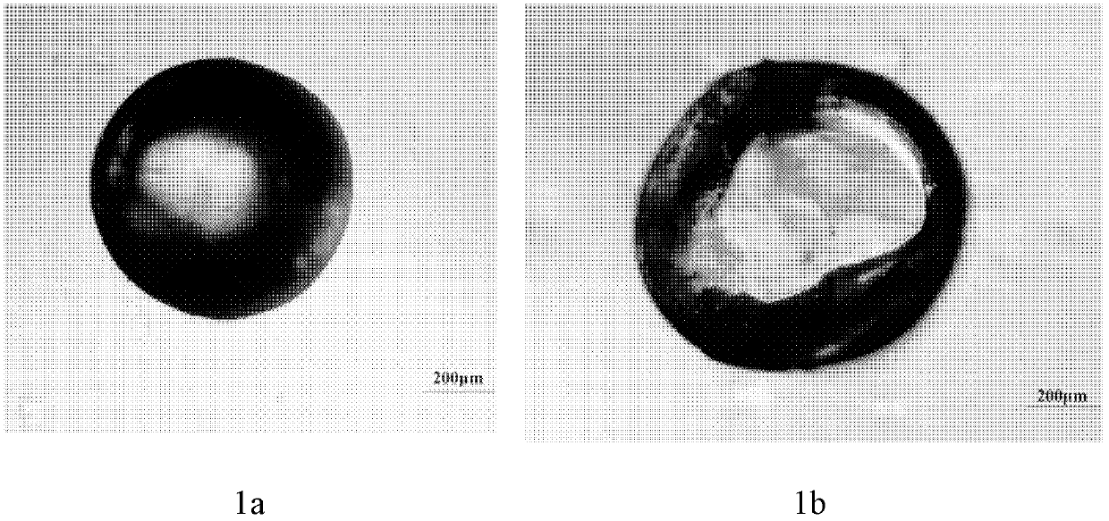
[0052] Embodiment 1: the preparation of polyvinyl alcohol iodized oil particle

[0053] Weigh 1 part of polyvinyl alcohol and prepare a 2% (w / v) polyvinyl alcohol solution. Add 10 parts of iodized oil into the polyvinyl alcohol solution and stir to form an emulsion. Take by weighing 4 parts of sodium sulfate, be mixed with 20% (w / v) sodium sulfate solution, add sodium sulfate solution in the emulsion, continue to stir, and make water bath heat up slowly, when temperature rises to the cloud point temperature under this condition At this time, add 5 parts of formaldehyde and 2 parts of dilute sulfuric acid, keep the temperature of the water bath, and cure for 24 hours. Stand for stratification, pour out the supernatant, filter and wash to obtain microcapsule embolism particles, the microscopic morphology of which is as follows: figure 1 shown.

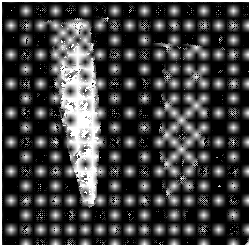
[0054] Determination of particle properties

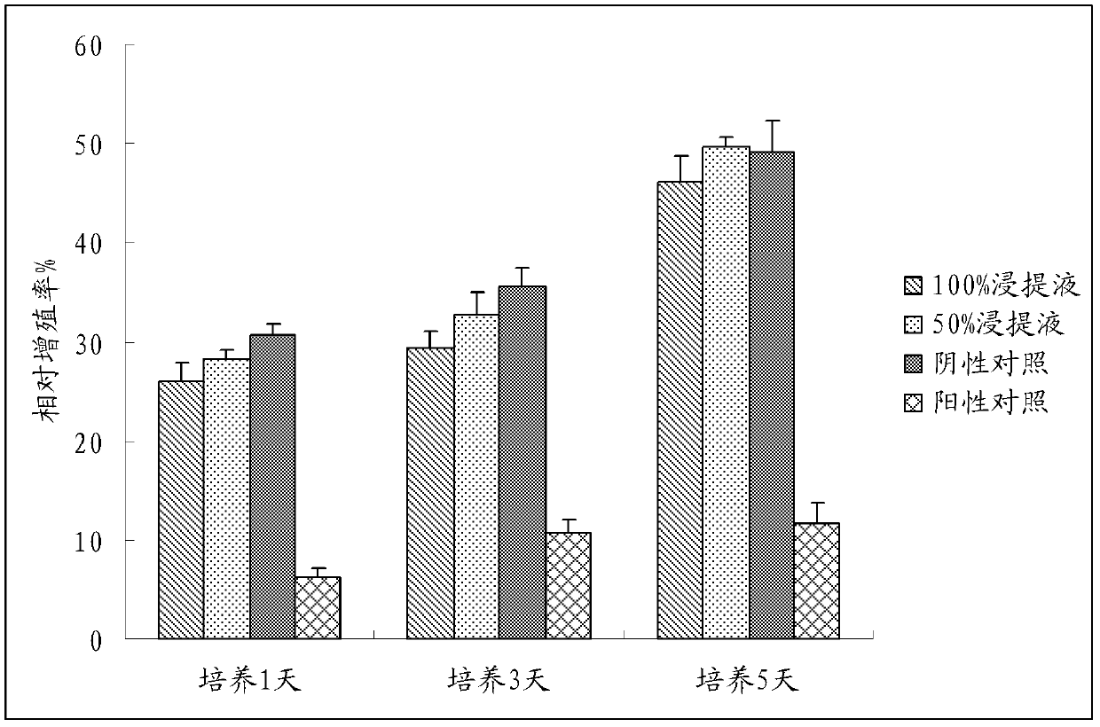
[0055] a. Cytocompatibility test

[0056] Use 10% fetal bovine serum containing doub...

Embodiment 2

[0073] Embodiment 2: the preparation of polyvinyl alcohol iodized oil particle

[0074] Weigh 1 part of polyvinyl alcohol to prepare a 1% (w / v) polyvinyl alcohol solution. Weigh 4 parts of sodium sulfate, prepare a 20% (w / v) sodium sulfate solution, and mix it with polyvinyl alcohol solution evenly. Add 8 parts of iodized oil to the above mixed solution, stir to form an emulsion, and slowly raise the temperature of the water bath, when the temperature rises to the cloud point temperature under this condition, add 5 parts of formaldehyde and 2 parts of dilute sulfuric acid, keep the water bath temperature, curing for 24 hours. Stand to separate layers, pour out the supernatant, filter and wash to obtain microcapsule embolism particles.

[0075] The cytocompatibility was measured by the same method as in Example 1, and the results showed that the cytocompatibility was good.

[0076] The developing effect under X-ray was measured by the same method as in Example 1, and the res...

Embodiment 3

[0079] Embodiment 3: the preparation of gelatin iodized oil particle

[0080] Weigh 1 part of gelatin and make a 5% (w / v) solution after swelling with distilled water. Under the condition of a water bath at 55°C, add 4 parts of iodized oil and stir at high speed to form an emulsion. Add 10% (w / v) acetic acid solution dropwise to adjust the pH to about 4.0. After observing the gelatin wrapped iodized oil under a microscope, add distilled water at 30°C to dilute, and stir to lower the temperature to room temperature. Add 0.4 parts of formaldehyde to solidify for 15 minutes under ice-water bath conditions, add dropwise 10% (w / v) sodium hydroxide solution, adjust the pH to 8-9, and continue to solidify for 4 hours. Stand to separate layers, pour out the supernatant, filter and wash.

[0081] The cytocompatibility was measured by the same method as in Example 1, and the results showed that the cytocompatibility was good.

[0082] The developing effect under X-ray was measured by...


PUM

PropertyMeasurementUnit
solubility (mass)aaaaaaaaaa
particle diameteraaaaaaaaaa

Abstract

The invention discloses embolization particles developable under X-rays. The particles contain a biocompatible material and iodized oil, and the iodized oil is coated by the biocompatible material to form microcapsule particles. The particles have high biocompatibility, can be monitored in embolization, and are convenient for checking the embolization effect after embolization, and the like; during entrapment of medicaments, the medicaments can be released slowly from the particles; relatively high medicament concentration can be kept for a long time on an embolization part; and compared with perfusion treatment, the embolization particles have the advantages: the toxic and side effects of medicaments on a whole body can be lowered, and treatment effect of embolization can be improved. Moreover, a preparation process of the embolization particles is simple, has low cost, and is suitable for large-scale industrial production.

Description

technical field [0001] The invention belongs to the field of interventional medicine, and in particular relates to X-ray-developable microparticles for embolism, a preparation method and application thereof. Background technique [0002] Interventional embolization therapy refers to the introduction of embolic agents into the human body through special precision instruments such as catheters and guide wires for local treatment under the guidance of medical imaging equipment. Embolization therapy has achieved good results in the treatment of uterine fibroids, liver cancer, kidney cancer, hemangioma, vascular malformation and hemostasis, and has become an alternative to partial surgical treatment. [0003] Among the various types of embolic agents, the application of particulate embolic agents is the most common. Ordinary embolic particles can be transmitted by X-rays, that is, there is no image under X-ray imaging equipment. On the contrary, embolic particles that can be vi...

Claims


Application Information

Patent Timeline
Patent Type & Authority Applications(China)
IPC IPC(8): A61L31/18A61L31/16A61L31/04A61L31/06A61K9/14A61K47/32A61K47/34A61K47/36A61K47/38A61K47/42A61P7/04A61P9/00A61P15/00A61P35/00
Inventor 范田园张苑
Owner HYGEA MEDICAL TECH CO LTD
Why Eureka
Try Eureka