Extraction and multiplication culture method and application of urine mesenchymal stem cells

A technique for stem cell expansion and culture, applied in the field of cell biology and regenerative medicine, can solve the problems of high cost, restrict wide application, invasive separation and extraction methods, etc., achieve good monitoring or tracking, and the method is simple and easy to implement The effect of amplifying power

Active Publication Date: 2013-02-13
上海艾棵颂生物科技有限公司 +1
View PDF3 Cites 24 Cited by
  • Summary
  • Abstract
  • Description
  • Claims
  • Application Information

AI Technical Summary

Problems solved by technology

Bone marrow-derived mesenchymal stem cells are the most studied and widely used adult stem cells, but the isolati

Method used


Image

  • Extraction and multiplication culture method and application of urine mesenchymal stem cells
  • Extraction and multiplication culture method and application of urine mesenchymal stem cells
  • Extraction and multiplication culture method and application of urine mesenchymal stem cells

Examples


Embodiment 1

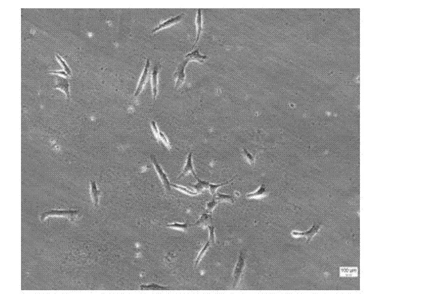
[0042] Isolation and extraction of urinary mesenchymal stem cells from clean midstream urine:

[0043] Under sterile conditions, take 200-250ml of clean middle urine from healthy volunteers. To prevent infection, add 5ml of double antibody containing penicillin 100U / ml + streptomycin 0.1mg / ml, and separate and extract immediately. Divide the clean urine into 50ml centrifuge tubes, centrifuge at 400g for 10min, and discard the supernatant. Wash twice by centrifugation at 400 g for 10 min with PBS. Take 100 μL of cell resuspension, use trypan blue to detect cell viability and count, adjust cell density, inoculate into a six-well plate containing serum-free medium, and place at 37°C in 5% CO 2 cultured in a humidified incubator.

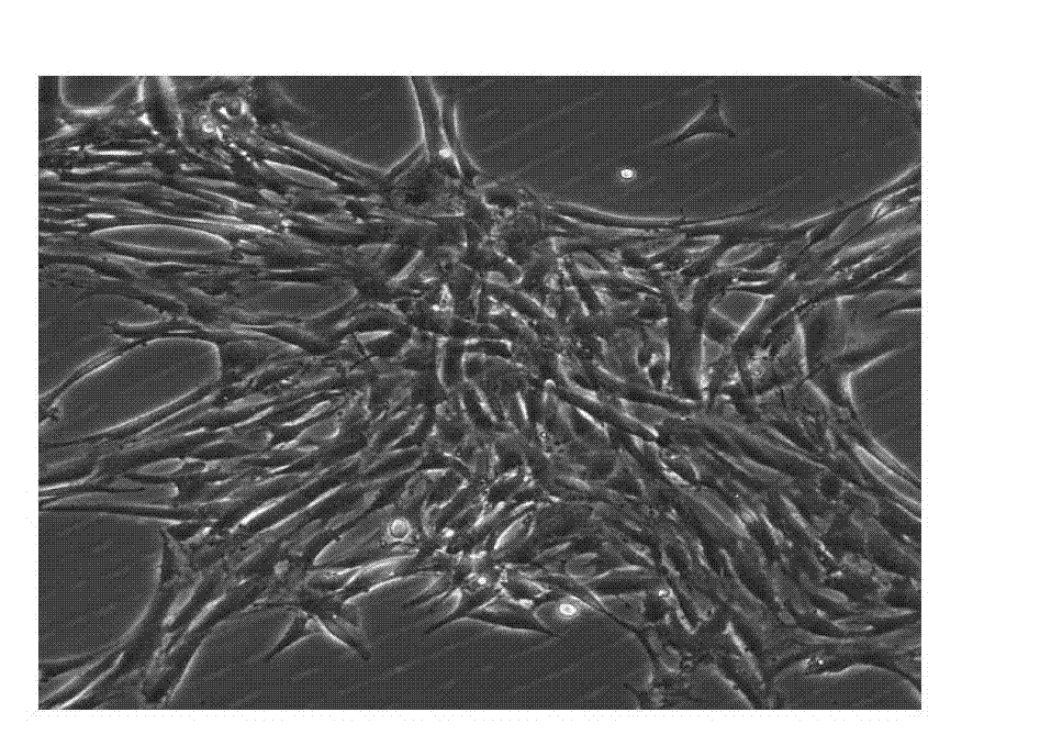
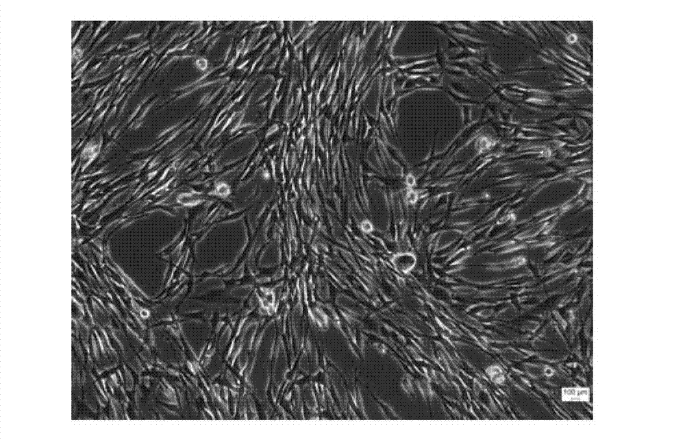
[0044] After 3-7 days of culture, observe under a phase-contrast microscope. If bacterial contamination is observed, discard it. If there is no contamination, continue the culture. A single cell can be found to adhere to the wall, and the position of ...

Embodiment 2

[0046] Differentiation of urinary mesenchymal stem cells into osteocytes:

[0047] (1) Take the fourth-generation culture of urine mesenchymal stem cells obtained in Example 1

[0048] (2) Induction culture medium containing 0.1 μmol / L of dexamethasone, 10 nmol / L of vitamin D, and 10 mmol / L of β-glycerol phosphate was added to the urinary mesenchymal stem cells for induction culture, and the medium was changed twice a week.

[0049] (3) Alizarin red staining was performed to observe calcium nodules after 21 days of induced differentiation, and RT-PCR was used to detect the expression of osteopontin, osteocalcin, and bone morphogenetic protein.

[0050] The results showed that a large number of calcium nodules appeared in the induced cells (see Figure 12 ), the expression levels of osteopontin, osteocalcin, bone morphogenetic protein and other genes were significantly increased, indicating that urinary mesenchymal stem cells can differentiate into osteoblasts.

Embodiment 3

[0052] Differentiation of urinary mesenchymal stem cells into chondrocytes:

[0053] (1) Take the fourth-generation culture of urine mesenchymal stem cells obtained in Example 1

[0054] (2) Add induction medium containing transforming growth factor β 110 ng / ml, insulin-like growth factor 100 ng / ml and dexamethasone 0.1 μmol / L to urinary mesenchymal stem cells

[0055] (3) After induction and culture for 28 days, the extracellular matrix of the cells was stained with Alcian blue, and the expression of type II collagen was detected by immunohistochemistry.

[0056] The results showed that most of the induced and cultured urinary mesenchymal stem cells were stained with Alcian blue, and the expression of type II collagen was enhanced, indicating that the urinary mesenchymal stem cells could differentiate into chondrocytes.


PUM


Abstract

The invention discloses an extraction and multiplication culture method and application of urine mesenchymal stem cells. The urine mesenchymal stem cells are separated from human urine, and are cultured and multiplied in a culture medium containing cell growth factors. The urine mesenchymal stem cells extracted and multiplication-cultured by the invention can repair various tissue and organ damage. The invention has the advantages that the sources of the cells are wide, the extraction and the multiplication culture are simple and feasible, and the popularization and the implementation are facilitated. The extraction method of the urine mesenchymal stem cells has the characteristics of no damage and wide sources, can induce and differentiate the urine mesenchymal stem cells into multiple cells, and has a wide application prospect in tissue engineering and regenerative medicine.

Description

technical field [0001] The invention relates to cell biology and regenerative medicine, in particular to a method and application for extracting, expanding and culturing urine mesenchymal stem cells. Background technique [0002] The development of regenerative medicine and tissue engineering technology has provided new means and methods for tissue regeneration and repair, but it still faces many challenges, among which the selection of suitable cell sources has always puzzled researchers. Currently, seed cells for regenerative medicine and tissue engineering mainly include: autologous or allogeneic tissue-specific cells, adult pluripotent stem cells, totipotent stem cells, and induced pluripotent stem cells. Autologous cell transplantation has the characteristics of high histocompatibility and safety, but its source is limited, the acquisition method is invasive, and it may cause dysfunction of the donor site. Allogeneic cell transplantation is easy to obtain and avoids su...

Claims


Application Information

Patent Timeline
IPC IPC(8): C12N5/0775C12N5/077C12N5/079C12N5/071A61L27/38
Inventor 汪泱关俊杰邓志锋张长青龚飞翔牛鑫田寿福胡斌郭尚春
Owner 上海艾棵颂生物科技有限公司
Why Eureka
Try Eureka