Method for clearing non-human animal skin heterologous antigen, and low immunogenicity non-human animal skin preparation method

An animal skin, immunogenic technology, applied in medical science, prosthesis, etc., can solve the problem of no clear indication of α-Gal antigen, and achieve the effect of low price, wide material source and high enzymatic activity.

Active Publication Date: 2013-10-16
FIELD OPERATION BLOOD TRANSFUSION INST OF PLA SCI ACAD OF MILITARY
View PDF9 Cites 6 Cited by
  • Summary
  • Abstract
  • Description
  • Claims
  • Application Information

AI Technical Summary

Problems solved by technology

The literature is silent on whether and how xenogeneic skin α-Gal antigens can be cleared

Method used


Image

  • Method for clearing non-human animal skin heterologous antigen, and low immunogenicity non-human animal skin preparation method
  • Method for clearing non-human animal skin heterologous antigen, and low immunogenicity non-human animal skin preparation method
  • Method for clearing non-human animal skin heterologous antigen, and low immunogenicity non-human animal skin preparation method

Examples


Embodiment 1

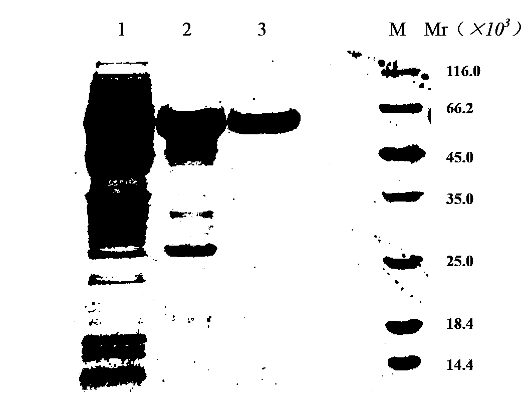
[0057] Embodiment 1, preparation recombinant α-galactosidase

[0058] Take the recombinant α-galactosidase strain pET22b-Gal / BL21(DE3) frozen at -70°C (see the Chinese patent document with publication number CN101845425A for the construction method), and put it on the LB plate (Amp + ) on the line to activate the strain, pick a single clone and culture overnight (10-12 hours) on the second day, and transfer to expand the culture to D on the third day 600 Add IPTG (final concentration: 0.1mM) to induce the expression of the target protein at 0.8, the induction time is 4 hours, collect the bacteria by centrifugation, and use Buffer A (20mM citric acid-disodium hydrogen phosphate buffer, 25mM NaCl, pH5.0) Suspend the cells, sonicate and lyse the cells, collect the supernatant by centrifugation (4°C, 9000r / min for 20min), and load the supernatant onto a cation chromatography column (Hitrap TM SP-HP), wash the column with 10 times the column volume of Buffer A, and use Buffer B (...

Embodiment 2

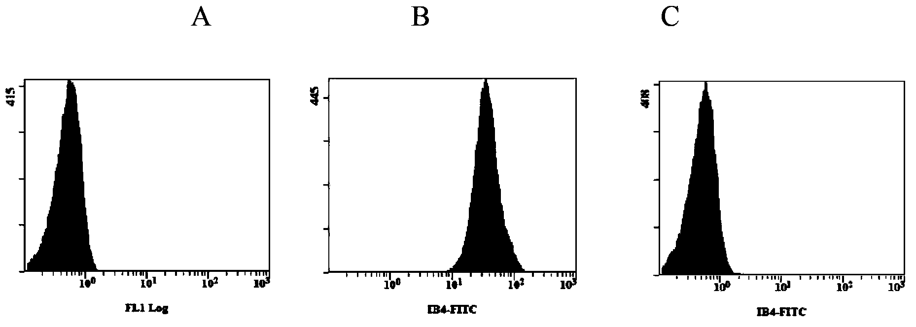
[0059]Embodiment 2, the detection of enzymatic hydrolysis experiment of porcine erythrocyte and α-Gal antigen clearance rate

[0060] In the experiment, the recombinant α-galactosidase can be prepared according to the method of Example 1; the enzymatic hydrolysis buffer is a glucose solution with a mass concentration of 5%.

[0061] Pig erythrocytes were washed twice with enzymatic hydrolysis buffer at a volume ratio of 1:1 and 1:4, so that the hematocrit was 40%; a recombinant α-galactosidase ( Prepared as in Example 1), slowly shake and incubate for 1 hour in an enzymatic hydrolysis reaction box at 26°C; then use PBS solution (recipe: each L contains 8g of NaCl, Na 2 HPO 4 1.44g, KCl0.2g, KH 2 PO 4 0.24g) wash red blood cells 4 times at a volume ratio of 1:4, add 1 / 2 volume of MAP solution (recipe: every L contains C 6 h 5 Na 3 o 7 .2H 2 O1.5g, C 6 h 8 o 7 .H 2 O0.2g, C 6 h 12 o 6 .H 2 O7.93g, NaH 2 PO 4 .2H 2 O0.94g, C 5 h 5 N 5 0.14g, NaCl4.97g, C 6 ...

Embodiment 3

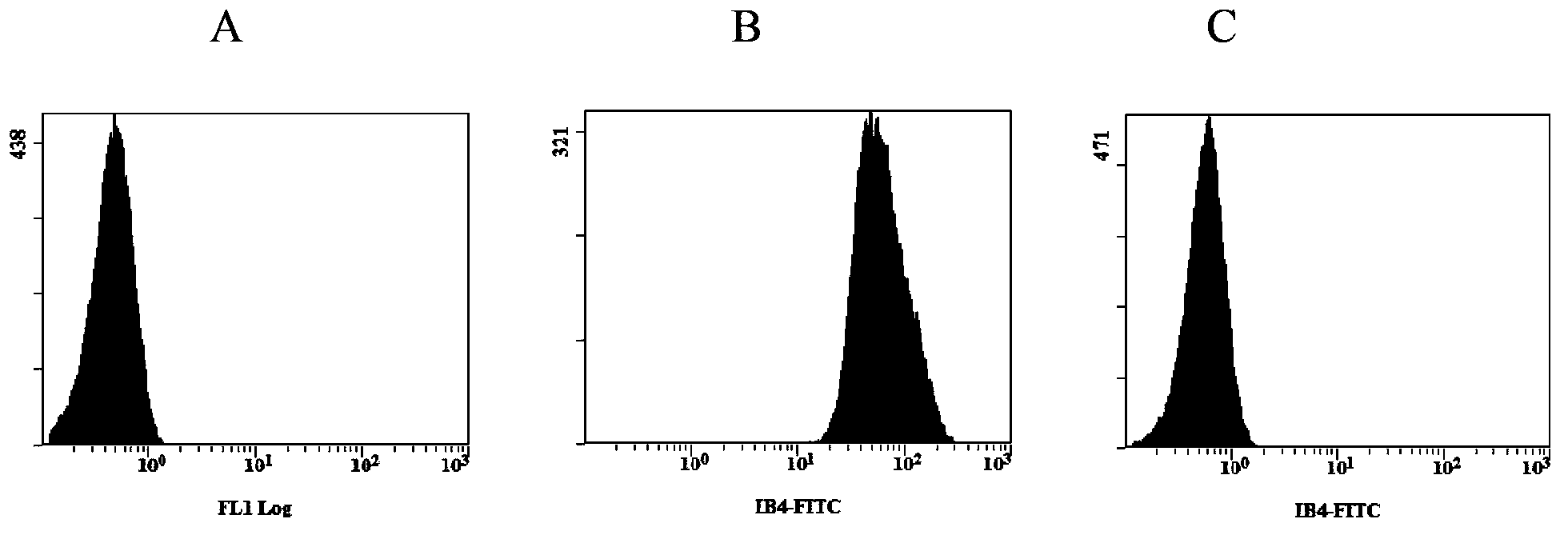
[0065] Embodiment 3, the detection of enzymatic hydrolysis experiment of bovine erythrocyte and α-Gal antigen clearance rate

[0066] The same operation as in Example 2, using bovine erythrocytes to carry out the enzymatic hydrolysis experiment.

[0067] The results of flow cytometry were as image 3 (A. blank (blank, unlabeled bovine erythrocytes), B. non-enzymolyzed bovine erythrocytes (positive control), C. enzymatically treated bovine erythrocytes), the test results show that the α-Gal antigen on the surface of bovine erythrocytes It can be cleared by α-galactosidase, and the clearance rate is greater than 99%.


PUM


Abstract

The present invention discloses a low immunogenicity non-human animal skin, a preparation method and applications thereof, wherein the low immunogenicity non-human animal skin is a non-human animal skin with clearing of heterologous antigen alpha-galactose (alpha-Gal) on the cell surface through a recombinant alpha-galactosidase enzymolysis treatment. The non-human animal skin has a characteristic of low immunogenicity, wherein the mainly reason is clearing of heterologous antigen alpha-Gal so as to prolong an occurrence time of an immunological rejection reaction after transplantation. In addition, the non-human animal skin can be provided for covering burning wound surfaces of burn patients, reducing immunological rejections during heterologous transplantation processes, and prolonging survival times of animal skins on burning wound surfaces so as to achieve purposes of wound surface healing promoting and scarring reducing.

Description

technical field [0001] The invention belongs to the field of biology, and relates to the preparation of heterogeneous skin, in particular to a method of treating heterogeneous skin with α-galactosidase to obtain non-human animal skin with low immunogenicity, and the skin can be used as a medical dressing for covering burn patients Burn wounds, promote wound healing, reduce scar formation. Background technique [0002] Burns are one of the common diseases in life, the incidence rate is about 1 in 1,000, and 1 in 10 of them need to be admitted to hospital for treatment. Burns are characterized by great damage, high risk and susceptibility to infection, shock and even death. Burned personnel in fires, especially those with large areas of burns, if the wounds are not promptly and effectively sealed, it will inevitably lead to internal environment disorder and invasion of a large number of pathogens, leading to shock, infection, multiple organ dysfunction and other severe diseas...

Claims


Application Information

Patent Timeline
Patent Type & Authority Applications(China)
IPC IPC(8): A61L27/36A61L27/60
Inventor 宫锋季守平高红伟李素波檀英霞鲍国强王颖丽
Owner FIELD OPERATION BLOOD TRANSFUSION INST OF PLA SCI ACAD OF MILITARY
Why Eureka
Try Eureka