Fluorescent glucose nano biosensor and preparation method thereof
A biosensor, glucose technology, applied in the field of biochemical analysis and quantitative research, to achieve the effect of high sensitivity, low toxicity and good stability
- Summary
- Abstract
- Description
- Claims
- Application Information
AI Technical Summary
Problems solved by technology
Method used
Image
Examples
Embodiment 1
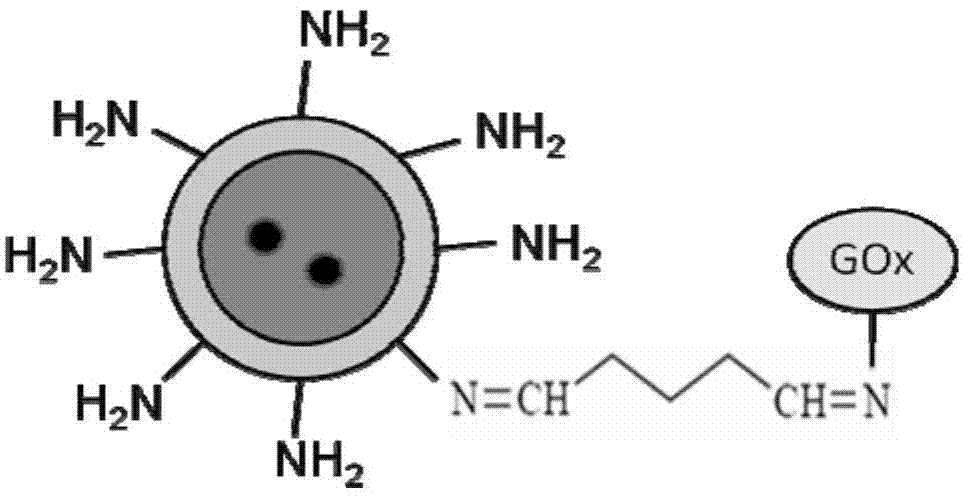
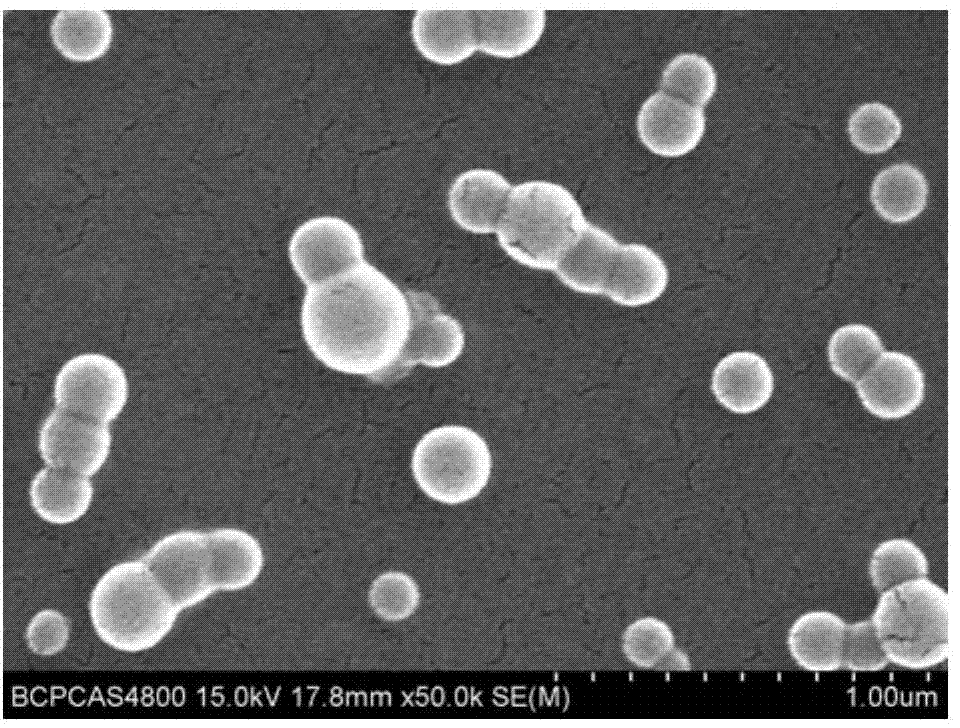
[0029] A fluorescent type glucose nanometer biosensor, which is composed of functionalized fluorescent nanoparticles, and has a structure such as figure 1 Shown. Firstly, a ratio fluorescent oxygen nanosensor coated with polylysine (PLL) shell layer was prepared by reprecipitation method: the inside is formed by hybridization of dodecyltrimethoxysilane (DTS) and polystyrene (PS) The core is doped with two fluorescent dye molecules (oxygen probe molecule platinum (Ⅱ) MESO-tetrakis (pentafluorobenzene) porphine (PtTFPP) and fluorescent reference molecule coumarin 6 (C6)); the exterior is PLL shell. The morphology and particle size of the oxygen nanosensor are as figure 2 As shown, the particle size is concentrated around 258.2nm. Then, based on the Schiff base reaction between primary amine and glutaraldehyde, glucose oxidase is coupled to the surface of the oxygen nanosensor, so that it can catalyze the oxidation of glucose. The specific coupling reaction principle is as follo...
Embodiment 2
[0036] PtTFPP, C6, PS and DTS were dissolved in tetrahydrofuran in a mass ratio of 2:2:46:50, and their total concentration in the solution was 0.2%. Then, use a micro-adjusting syringe to take 500 μL of the solution, and quickly inject it into deionized water with a PLL concentration of 0.02 mg / ml with a pH of 9 (adjust the pH with ammonia water) under ultrasonic vibration, and the resulting suspension is ultrasonically After shaking for 2 hours, followed by dialysis in deionized water for 24 hours, PLL-coated core-shell structured nanoparticles were obtained, which are sensitive to oxygen, and the fluorescence intensity decreases with increasing oxygen concentration. This is the fluorescent nano oxygen sensor.
[0037] In the fluorescent nano-oxygen sensor prepared above, that is, the PLL-coated nanoparticles, first add the same amount of glutaraldehyde as the PLL substance, shake for 5 minutes at 25°C, and mix it uniformly; then add glucose Oxidase (GO x ) In the aqueous solu...
Embodiment 3
[0043] 1) Dissolve platinum (II) MESO-tetrakis (pentafluorobenzene) porphine, coumarin 6, polystyrene and dodecyltrimethoxysilane with a mass ratio of 1:2:47:50 in tetrahydrofuran , The total concentration in the solution is 0.1%;
[0044] 2) Prepare a deionized aqueous solution of polylysine with a concentration of 0.02 mg / ml, and adjust the pH of the deionized aqueous solution of polylysine to 8 with ammonia;
[0045] 3) Take 300 μL of the tetrahydrofuran solution described in step 1) and inject it into the deionized aqueous solution of polylysine described in step 2) under ultrasonic vibration to obtain a suspension. After ultrasonic vibration for 1 hour, it is then deionized After 12 hours of dialysis in water, fluorescent oxygen nanoparticles with core-shell structure are obtained, and the particle size is about 240nm;
[0046] 4) In step 3) the fluorescent oxygen nanoparticles with core-shell structure, first add glutaraldehyde in the amount of polylysine and other substances,...
PUM
| Property | Measurement | Unit |
|---|---|---|
| particle diameter | aaaaa | aaaaa |
| particle diameter | aaaaa | aaaaa |
| particle diameter | aaaaa | aaaaa |
Abstract
Description
Claims
Application Information
 - R&D
- Intellectual Property
- Life Sciences
- Materials
- Tech Scout
- Unparalleled Data Quality
- Higher Quality Content
- 60% Fewer Hallucinations
Browse by: Latest US Patents, China's latest patents, Technical Efficacy Thesaurus, Application Domain, Technology Topic, Popular Technical Reports.
© 2025 PatSnap. All rights reserved.Legal|Privacy policy|Modern Slavery Act Transparency Statement|Sitemap|About US| Contact US: help@patsnap.com


