Method for preparing three-dimensional porous stent composite layer

A three-dimensional porous and composite layer technology, applied in medical science, prosthesis, etc., can solve the problems of cumbersome procedures and inability to effectively guarantee the penetration of pores, and achieve the effect of low production cost and good biocompatibility

Inactive Publication Date: 2014-04-02
SOUTHWEST JIAOTONG UNIV
View PDF7 Cites 14 Cited by
  • Summary
  • Abstract
  • Description
  • Claims
  • Application Information

AI Technical Summary

Problems solved by technology

From the synthesis of powder to the output of the scaffold, the process is relatively cumbersome. In order to obtain high mechani

Method used


Image

  • Method for preparing three-dimensional porous stent composite layer
  • Method for preparing three-dimensional porous stent composite layer
  • Method for preparing three-dimensional porous stent composite layer

Examples


Example Embodiment

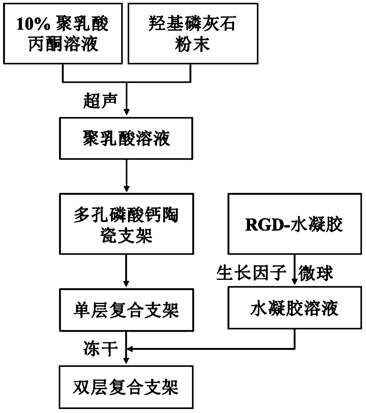
[0026] Example 1: Weigh 2.0 g of polylactic acid and dissolve it in 20 mL to prepare a solution with a mass volume ratio of 10%, then add 10% mass fraction of hydroxyapatite dry powder, and ultrasonically disperse and discard the underlying precipitate. The spare three-dimensional porous scaffold was placed in the solution, taken out after one minute and dried; the 10% arginine-glycine-aspartic acid sequence (RGD) graft was prepared with a pH=7.4 phosphate buffer solution. of oxidized sodium alginate solution and 2.5% N-succinyl chitosan solution. After sterilization, 9 mL of N-succinyl chitosan solution and 1 mL of RGD-grafted oxidized sodium alginate solution were mixed, and quickly stirred uniformly. The three-dimensional porous scaffold previously compounded with polylactic acid is put into the mixed solution, and after being quickly and uniformly coated, the excess hydrogel on the surface is removed; after freeze-drying, a three-dimensional porous scaffold compound layer ...

Example Embodiment

[0027] Example 2: Weigh 2.0g of polylactic acid and dissolve it in 20mL to prepare a solution with a mass-to-volume ratio of 10%, add 10% of hydroxyapatite dry powder by mass, and after ultrasonic dispersion, discard the underlying precipitate, and place the spare three-dimensional porous support into the solution. In this solution, take it out after one minute and spin dry; prepare 10% RGD-grafted oxidized sodium alginate solution and 2.5% N-succinyl chitosan solution with pH=7.4 phosphate buffer solution. After sterilization, 9 mL of N-succinyl chitosan solution and 1 mL of RGD grafted oxidized sodium alginate solution were mixed, and VEGF at a concentration of 10 ng / mL was added. 165 (vascular endothelial growth factor), BMP-2 (cell differentiation growth factor) at a concentration of 10ng / mL, and 10mg sodium alginate microspheres loaded with TGF-beat1 (transforming growth factor) and quickly stirred evenly. The three-dimensional porous scaffold is put into the mixed soluti...

Example Embodiment

[0028] Example 3: Weigh 2.0 g of polylactic acid and dissolve it in 20 mL to prepare a solution with a mass-to-volume ratio of 10%, add hydroxyapatite dry powder with a mass fraction of 10%, discard the bottom precipitate after ultrasonic dispersion, and place the porous scaffold in the dispersion. After one minute, take out and spin dry; prepare 10% RGD-grafted oxidized sodium alginate solution and 2.5% N-succinyl chitosan solution with pH=7.4 phosphate buffer solution, sterilize Then, 8 mL of N-succinyl chitosan solution was mixed with 2 mL of RGD grafted oxidized sodium alginate solution, and VEGF at a concentration of 10 ng / mL was added. 165 , 10ng / mL concentration of BMP-2 and 10mg were loaded into TGF-beat1 sodium alginate microspheres and quickly stirred uniformly. The three-dimensional porous scaffold previously compounded with polylactic acid was put into the mixed solution, and after rapid coating uniformly, the surface was removed. Excess hydrogel; freeze-drying to ...


PUM


Abstract

The invention discloses a method for preparing a three-dimensional porous stent composite layer, and belongs to the technical field of biomaterials. In order to prevent inflammatory reaction caused by drop of scraps of a stent due to a dynamic action, a coating film is required on the surface of a porous ceramic stent, and addition of hydroxyapatite powder fills up a voidness defect of the surface layer of the stent so as to improve the strength of the stent; a porous calcium phosphate ceramic stent is compounded by a polylactic acid solution added with hydroxyapatite and a growth factor-loaded hydrogel solution; growth factors loaded in the hydrogel can be slowly released to a human part needing the growth factors under the joint action of own diffusion and slow degradation of polymeric hydrogel so as to promote vascular endothelial growth of a tissue and proliferation and differentiation of an osteoblast; and the stent is gradually degraded and eventually transformed into a bone; and the method is mainly used for preparing the three-dimensional porous stent composite layer.

Description

technical field [0001] The invention belongs to the technical field of biological materials, in particular to a composite layer of a bone repair material and a bone tissue engineering material and a preparation method thereof. Background technique [0002] Calcium phosphate ceramics have good biocompatibility, bioactivity, osteoinductivity, and degradability due to their similarity to the inorganic components of the human body's hard tissues. bonding, and is widely used as a bone repair material. In clinical practice, they can be widely used for the repair of bone defects in non-load-bearing and load-bearing parts, so good mechanical properties are a basic requirement of ceramic scaffolds. However, the research results show that dense calcium phosphate ceramics cannot effectively induce the formation of new bone tissue, and only calcium phosphate ceramics with porous structure have osteoinductive ability. Conducive to the growth of the organization. Based on this, there h...

Claims


Application Information

Patent Timeline
IPC IPC(8): A61L27/46A61L27/48A61L27/56A61L27/54A61L27/58
Inventor 汪建新闫浩然陈太军刘霞翁杰冯波鲁雄周绍兵屈树新段可卢晓英智伟
Owner SOUTHWEST JIAOTONG UNIV
Why Eureka
Try Eureka