

Method for detecting asbestos
一种检测方法、石棉的技术,应用在测量装置、颜色/光谱特性测量、仪器等方向,能够解决无法快速检测石棉、无法应对石棉风险、难多个处理等问题,达到简便且高精度检测的效果
- Summary
- Abstract
- Description
- Claims
- Application Information
AI Technical Summary
Problems solved by technology
Method used
Image

Examples
Embodiment 1
[0184] [Example 1: Asbestos binding protein H-NS 60-90 Preparation and fluorescent labeling]
[0185] The biotin-streptavidin interaction is used to combine the biotin-modified asbestos-binding protein with fluorescently labeled streptavidin to produce fluorescently-labeled asbestos-binding protein.
[0186] First, make biotinylated asbestos binding protein. Using protein expression vector pET21-b (Novagen) as a template, oligonucleotide primer P1 (GCTCAGAAAATCGAATGGCACGAACACCACCACCACCACCACTGAACTA: SEQ ID NO:1) and oligonucleotide primer P2 (CTCGAAGATGTCGTTCAGACCGCCACCCTCGAGTGCGGCCGCAAGCTTGTC: SEQ ID NO: 2) were used for reverse PCR , Thus insert the biotinylated tail before the HisTag of pET21-b. The reverse PCR reaction was performed using KOD-plus-mutagenic kit (TOYOBO) according to the company's protocol. The biotinylated protein expression vector was named "pET21-AviTag-C".
[0187] Then, using the genomic DNA of Escherichia coli K-12 (Escherichia coli K12, ATCC700926) as a t...
Embodiment 2
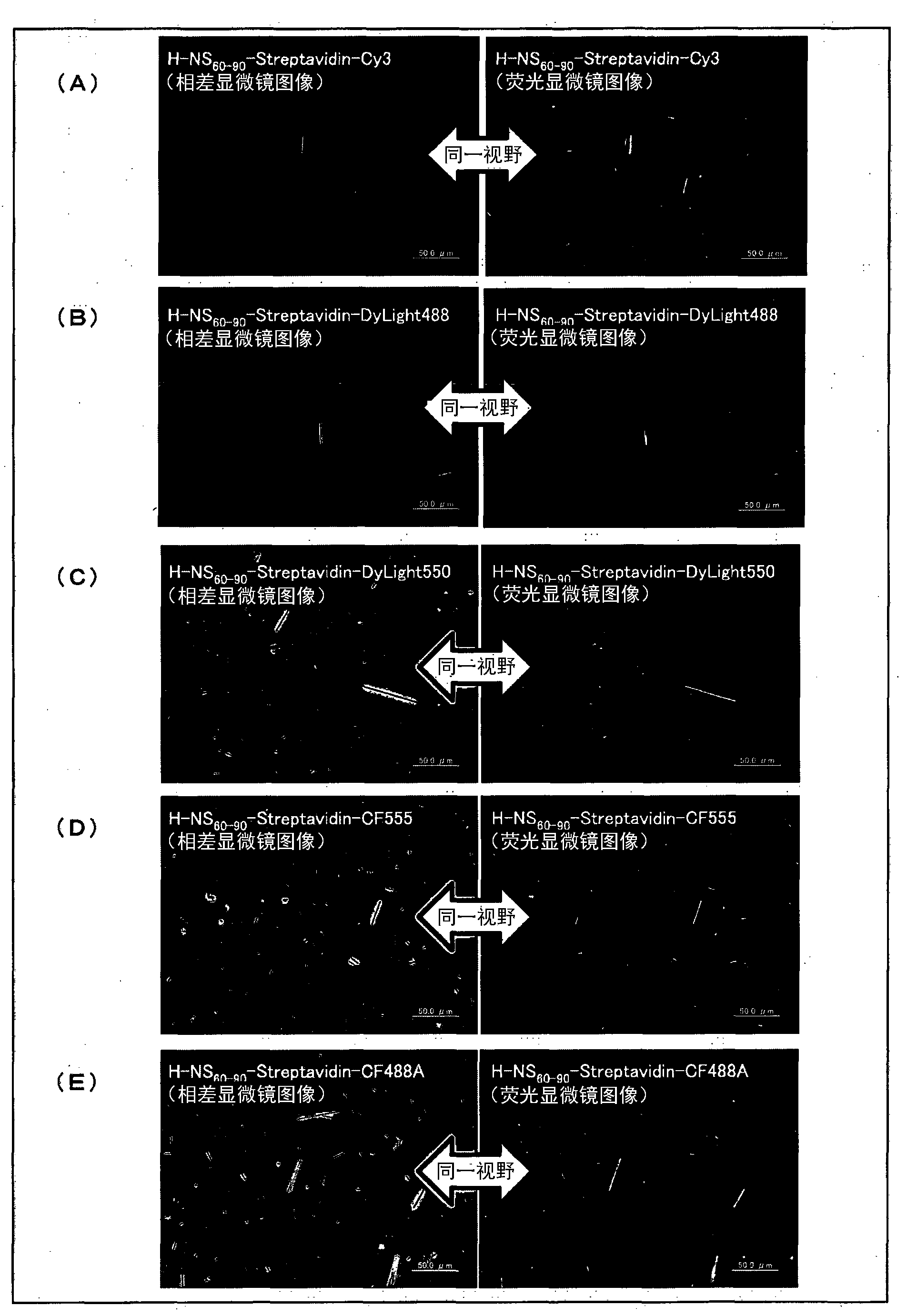
[0193] [Example 2: Asbestos Microscopic Observation by Phase Contrast Fluorescence Method (1)]
[0194] Cut the membrane filter containing asbestos amosite (thea asbestos) (JAWE231) as the test substance into 1 / 8 pieces, with the collecting side facing up, add 20μl of buffer C [0.3M phosphate buffer three times Solution (pH8.0), 0.3M NaCl, 0.5% Tween80 (registered trademark)].
[0195] Then, add 5 times 20μl of H-NS containing fluorescent protein 60-90 -Streptavidin-Cy3 (12.5nM), H-NS 60-90 -Streptavidin-DyLight488 (50nM), H-NS 60-90 -Streptavidin-DyLight550 (12.5nM), H-NS 60-90 -Streptavidin-CF555 (12.5nM) or H-NS 60-90 -Streptavidin-CF488A (50nM) buffer C to bind fluorescent protein with amosite. After that, 20 μl of buffer C was added dropwise 3 times to remove unbound fluorescent protein. Finally, add 20 μl of water three times to remove the surfactant or salt from the buffer.
[0196] Next, place the filter collection surface on a glass slide (Mitsunami, MICRO SLIDE GLASS, 1 m...
Embodiment 3
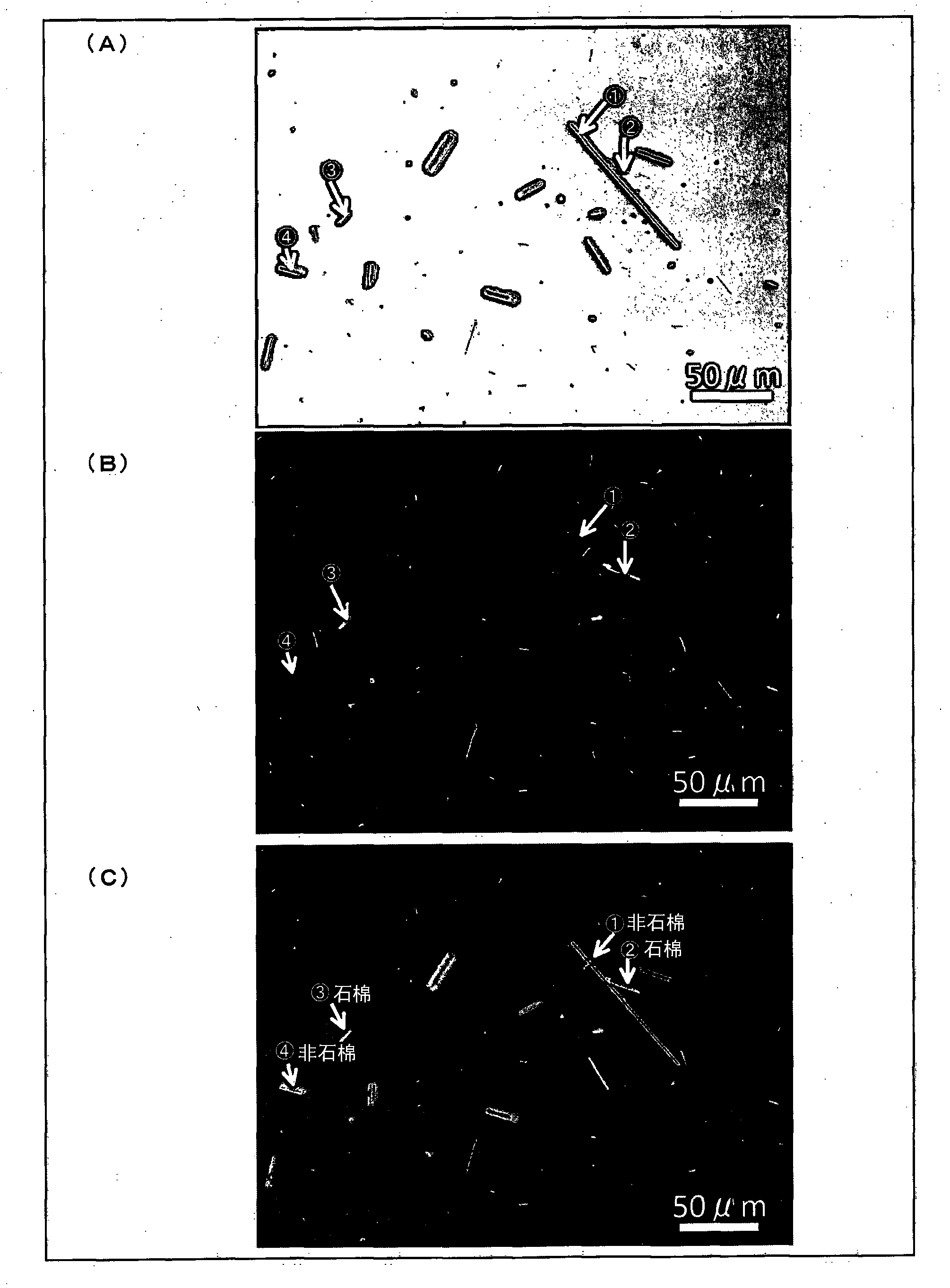
[0202] [Example 3: Asbestos Microscopic Observation by Phase Contrast Fluorescence Method (2)]
[0203] In addition to the use of membrane filters that capture amosite (thea asbestos) (JAWE231) and rock wool (JFM standard fiber samples) as the test objects, and only use "H-NS 60-90 -Streptavidin-Cy3" was used as a fluorescent protein, and a specimen was prepared by the same method as in Example 2.
[0204] Observe the obtained specimen with a phase contrast / fluorescence microscope (Epi-fluorescence microscope BX-60, manufactured by Olympus). First, observe with a phase contrast microscope to confirm the fibrous material, then switch the optical path to fluorescence mode and observe the same field of view.
[0205] figure 2 It is a diagram showing the result of microscopic observation of asbestos by phase contrast fluorescence method. figure 2 (A) represents the phase contrast microscope image, (B) represents the fluorescence microscope image in the same field as the phase contrast ...
PUM
 Abstract
Description
Claims
Application Information