Self-powered microtubule-kinesin transport system and preparation method thereof
A technology of kinesin and self-supply energy, which is applied in chemical instruments and methods, animal/human peptides, material inspection products, etc. It can solve the problems of activity reduction, kinesin and microtubule protein damage, cumbersome operation steps, etc.
- Summary
- Abstract
- Description
- Claims
- Application Information
AI Technical Summary
Problems solved by technology
Method used
Image
Examples
preparation example Construction
[0035] The preparation method of the silanized fluid cell is the same as the above method for preparing the common fluid cell, except that the slide glass and the cover glass need to be silanized in advance. Preparation of aminosilanized coverslips: 20 μl of 3-aminopropyltriethoxysilane (5 wt %) in acetone was dropped onto a clean slide and immediately covered with another clean slide. After 5 min, the slides were rinsed with water and dried with liquid nitrogen. This results in two aminosilanized coverslips. Prepare a fluid cell with the 2 coverslips described above.
[0036] In the following examples, the preparation method of the rhodamine-labeled biotin-modified microtubules used is: rhodamine-labeled microtubules, biotin-modified microtubules and unmodified microtubules (volume ratio 1:1:2 ) mixed well and dispersed in 4mM MgCl 2 , 1mM GTP (guanosine triphosphate) and 5wt% DMSO in the BRB80 buffer solution, the total protein concentration was 2.5mg / ml; then placed in a...
Embodiment 1
[0039] Example 1, preparation of microtubule-kinesin transport system
[0040] (1) Preparation of creatine kinase (CPK) microspheres
[0041] Take Na 2 CO 3 Water and CPK aqueous solution were placed in a round bottom flask, and then an equal volume of CaCl was quickly added at one time 2 Put the aqueous solution in a flask, stir it for 20 seconds immediately, let it stand for about 2 minutes, take the precipitate, centrifuge it, and wash it with water three times to obtain CPK microspheres.
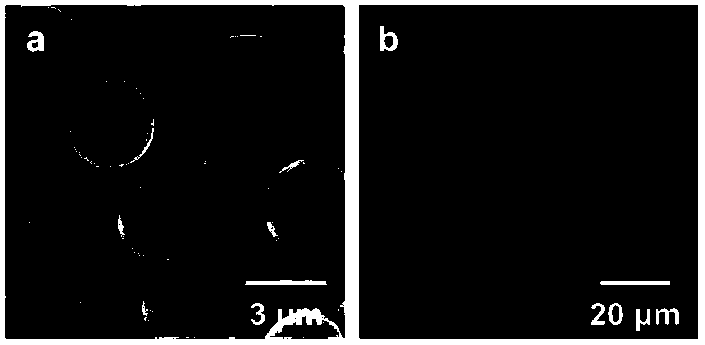
[0042]The scanning electron micrographs of the CPK microspheres prepared above are as follows: figure 1 As shown in (a), the laser confocal micrograph is as follows figure 1 As shown in (b), it can be seen from this figure that the particle size of the CPK microspheres prepared in this embodiment is 3.6 μm.
[0043] (2) Preparation of Microtubule-Creatine Kinase (MT-CPK) Microsphere Complex
[0044] The creatine kinase microspheres prepared in step (1) were dispersed into polyallyl...
Embodiment 2
[0051] Example 2, preparation of microtubule-kinesin transport system
[0052] The steps and parameters of this embodiment are basically the same as in Example 1, the difference is that when preparing creatine kinase microspheres, the Na 2 CO 3 Aqueous solution or CaCl 2 A drug model (fluorescein isothiocyanate-dextran (FITC-Dextran)) was added to the aqueous solution, and creatine kinase-drug composite microspheres were obtained by co-precipitation. Therefore, the composite microspheres prepared in this example can not only continuously provide ATP for the microtubule-kinesin transport system, but also serve as a carrier to transport drugs or other substances.
PUM
| Property | Measurement | Unit |
|---|---|---|
| Particle size | aaaaa | aaaaa |
| Particle size | aaaaa | aaaaa |
| Particle size | aaaaa | aaaaa |
Abstract
Description
Claims
Application Information
 - R&D
- Intellectual Property
- Life Sciences
- Materials
- Tech Scout
- Unparalleled Data Quality
- Higher Quality Content
- 60% Fewer Hallucinations
Browse by: Latest US Patents, China's latest patents, Technical Efficacy Thesaurus, Application Domain, Technology Topic, Popular Technical Reports.
© 2025 PatSnap. All rights reserved.Legal|Privacy policy|Modern Slavery Act Transparency Statement|Sitemap|About US| Contact US: help@patsnap.com


