Application of radioisotope-labeled monoanthranuclear anthraquinone compounds in the preparation of drugs for detecting myocardial activity
A radioactive isotope, monoanthranuclear anthraquinone technology, applied in the field of medicine, can solve problems such as wrong use of treatment methods, inability to accurately judge the state of heart cells, and delays in disease progression
- Summary
- Abstract
- Description
- Claims
- Application Information
AI Technical Summary
Problems solved by technology
Method used
Image
Examples
Embodiment 1
[0019] Example 1 Preparation of Myocardial Infarction Rat Model
[0020] SD rats with a body weight of 200-300 g were intraperitoneally injected with 10% chloral hydrate (0.3 mL / 100 g). After anesthesia, the rats were fixed on their backs on the mouse table, connected to a small animal ventilator through orotracheal intubation, the respiratory rate was 60-80 times / min, the respiratory ratio was 1 / 1, and the tidal volume was 4mL / 100g. After disinfecting the skin with povidone iodine, the chest was opened along the 3rd and 4th intercostal spaces along the left sternum to expose the heart, the pericardium was peeled off, a thread was threaded parallel to the left atrial appendage at the interventricular groove, and the left anterior descending coronary artery was ligated. After ligation, the air in the chest cavity was pumped out, the negative pressure in the chest cavity was restored, the chest cavity was quickly sutured, the endotracheal tube was withdrawn, and 160,000 U of pen...
Embodiment 2
[0021] Example 2 Preparation of iodine-131 labeled rhein
[0022] Weigh 0.4 mg rhein, dissolve it in 182.8 μl DMSO, and shake it well to obtain a 2.2 mg / ml rhein DMSO solution. Add 182.8 μl of rhein DMSO solution with a concentration of 2.2 mg / ml into a coated tube with an Iodogen content of 40 μg, add 45.7 μl of 17500 μCi Na iodine-131 solution, and then add 15 μl of PBS, shake well, and place in a water bath at 45°C The reaction was heated for about 90 minutes. After the reaction was terminated, the labeling rate was measured by TLC method. The labeling rate was greater than 95%, indicating that the labeling was successful. Labeling rate measurement method: the reaction solution was determined by paper chromatography, Whatman filter paper was used as the carrier, and 0.1mol / L HCl was used as the mobile phase for development. Labeling rate was measured by paper chromatography. Free 131I was distributed at the front of the solvent, while 131I-labeled rhein remained at the ori...
Embodiment 3
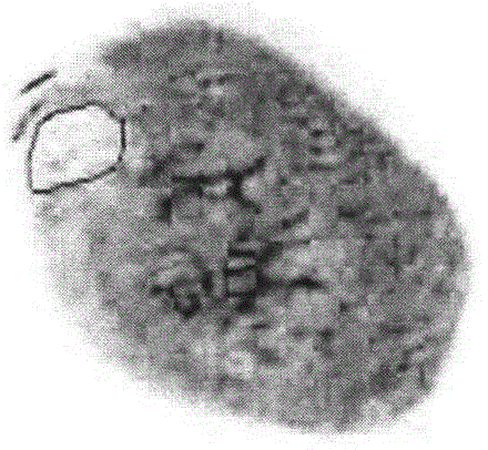
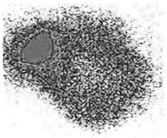
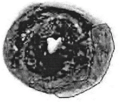
[0023] Example 3 Distribution of iodine-131-labeled rhein on rat model of myocardial infarction
[0024] The iodine-131 labeled rhein solution prepared in Example 2 was diluted with PEG 400-propylene glycol (1:1). Six myocardial infarction model rats were taken, and each rat was intravenously injected with 100 μCi of iodine-131-labeled rhein solution (radiochemical purity: 90%). After 12 hours, the model rats were euthanized, and various organs (thyroid, kidney, liver, spleen, lung, normal myocardium, infarcted myocardium, small intestine, stomach, muscle and fur, etc.) were taken, weighed separately, and radioactivity was measured with a gamma counter. , after decay correction, the results were expressed as the radioactive uptake per gram of organ or tissue as a percentage of the total injected dose (%ID / g).
[0025] After iodine-131 labeled rhein for 12 hours, the distribution value in necrotic myocardium was 0.78%ID / g, and that in normal myocardium was 0.15%ID / g, and the d...
PUM
 Abstract
Description
Claims
Application Information
 - R&D
- Intellectual Property
- Life Sciences
- Materials
- Tech Scout
- Unparalleled Data Quality
- Higher Quality Content
- 60% Fewer Hallucinations
Browse by: Latest US Patents, China's latest patents, Technical Efficacy Thesaurus, Application Domain, Technology Topic, Popular Technical Reports.
© 2025 PatSnap. All rights reserved.Legal|Privacy policy|Modern Slavery Act Transparency Statement|Sitemap|About US| Contact US: help@patsnap.com


