
Preparation method of tissue repair material

A tissue repair and membrane tissue technology, applied in the field of tissue engineering medical biomaterials, can solve problems affecting repair effect and functional recovery, easy to shrink, affect tissue recovery effect, etc., to achieve high biosafety and biocompatibility , small changes in mechanical properties and degradability, and the effect of reducing the risk of organic solvent residues

Active Publication Date: 2015-04-22
SHAANXI BIO REGENERATIVE MEDICINE CO LTD
View PDF10 Cites 36 Cited by
  • Summary
  • Abstract
  • Description
  • Claims
  • Application Information

AI Technical Summary

Problems solved by technology

[0006] Clinical studies have found that in some surgical operations, when the thickness of the material is too thin to fully fill the height of the damaged part, it will cause the repaired part to be sunken or the surface will be uneven, which will seriously affect its repair effect and functional recovery
In addition, during clinical use, thinner materials tend to shrink due to rehydration or absorption of tissue fluid, and adhere to surgical equipment, causing inconvenience to surgical operations
However, the thickness of biomaterials is limited by the raw materials themselves, such as pericardium, peritoneum, and bladder membrane, which are only 0.1-0.4 mm thick; It will inevitably lead to increased cost and reduced output, and also brings the risk of material delamination during use, which will affect the recovery effect of the tissue

Method used


Image

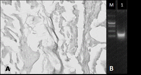
  • Preparation method of tissue repair material
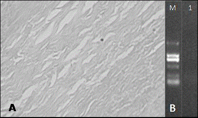
  • Preparation method of tissue repair material
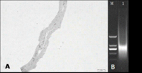
  • Preparation method of tissue repair material

Examples


Embodiment 1

[0041] Embodiment 1, Preparation of tissue repair materials from porcine peritoneum

[0042] Step 1. Pre-treatment: flatten the pig peritoneum on a flat plate, tear off a large piece of fat, remove the residual fat and connective tissue, wash with water until bloodless, and obtain the pig peritoneum material;

[0043] Step 2. Degreasing: Place the obtained membrane material in a mixed solution of methanol and chloroform (volume ratio 1:2), shake and degrease for 10 hours, then change the liquid, repeat the degreasing 3 times, and wash with water until odorless;

[0044] Step 3. Disinfection: Soak the degreased membrane material in an ethanol solution with a volume concentration of 75% for 3 hours, and wash it with water until it is odorless;

[0045] Step 4, decellularization: place the sterilized membrane material in a decellularized hypertonic solution containing 3M NaCl and 0.25 M NaOH, shake for 30 minutes, then transfer to water and shake for 30 minutes, and repeat this...

Embodiment 2

[0049] Embodiment 2, Preparation of tissue repair materials from sheep bladder matrix membrane

[0050] Step 1. Pretreatment: flatten the sheep bladder on a flat plate, separate the matrix membrane, wash with water until bloodless, and obtain the sheep bladder matrix membrane material;

[0051] Step 2, degreasing: place the obtained membrane material in acetone, shake and degrease for 2 hours, then change the liquid, continue shaking and degreasing for 4 hours, then wash with water until odorless;

[0052] Step 3. Disinfection: Soak the degreased membrane material in an ethanol solution with a volume concentration of 75% for 2 hours, and wash it with water until it is odorless;

[0053] Step 4, decellularization: place the sterilized membrane material in a decellularized hypertonic solution containing 1M NaCl and 0.2M HCl, shake for 15 minutes, then transfer to water and shake for 15 minutes, repeat this process 3 times to complete decellularization;

[0054] Step 5, swelli...

Embodiment 3

[0057] Embodiment 3, Preparation of tissue repair materials from bovine pericardium

[0058] Step 1, pre-treatment: flatten the bovine pericardium on a flat plate, tear off a large piece of fat on it, remove residual fat and connective tissue, wash with water until bloodless, and obtain the bovine pericardium material;

[0059] Step 2, degreasing: place the obtained membrane material in isopropanol, shake and degrease for 4 hours, and then change the liquid, then repeat the shaking and degreasing twice for 10 hours and 4 hours respectively, and wash with water until odorless;

[0060] Step 3. Disinfection: Soak the degreased membrane material in an ethanol solution with a volume concentration of 70% for 4 hours, and wash it with water until it is odorless;

[0061] Step 4, decellularization: place the sterilized membrane material in a decellularized hypertonic solution containing 5M NaCl and 1M NaOH, shake for 1 hour, then transfer to water and shake for 20 minutes, and repe...


PUM


Abstract

The invention relates to a preparation method of a tissue repair material. The preparation method comprises the following steps: carrying out pre-treatment; vibrating in an organic solvent to degrease; deactivating a virus with an ethanol solution; removing cells by high and low osmotic solutions; swelling in an acidic or alkaline liquid; and quickly lyophilizing and sterilizing. The thickness of the tissue repair material is increased by 2-4 times, and the material is loose and porous, small in change of mechanical property and degradation property and good in cell removal effect and has relatively high biosafety and biocompatibility. Due to the structural characteristics of proper thickness and looseness and porosity, the material is suitable for damage repair for filling damaged parts required by repair such as cartilage injury, skin and soft tissue defect, gingival recession and the like. Animal experiments verify that the repaired region after eight weeks has no depressed phenomena and is fully replaced by regenerated tissues. Moreover, the regenerated tissue and autologous tissue are healed well and are free of separating phenomenon, which verifies that the material can provide sufficient three-dimensional space for cell proliferation and crawl, so that the repaired region has good integration degree and filling degree, thereby realizing tissue repair and regeneration.

Description

Technical field [0001] The present invention is a technology field of organizing engineering and medical biological materials, which involves a method of preparation of organizational repair materials. Background technique [0002] Organization and organ loss or dysfunction are one of the most frequent and serious issues facing human health.According to statistics, millions of people in the United States suffer from various organizations, organ loss or dysfunction every year, and 8 million surgery is required each year, with an annual cost of more than 40 billion US dollars.In recent years, with the development of organizational engineering and regeneration medicine, the repair and treatment of tissue and organs has made many breakthroughs. Among them, the heterogeneous dehydrated cell biofilm material has gained more attention due to its unique natural structure and the degradation performance of the regeneration synchronization with the tissue regeneration.EssenceMany research ...

Claims


Application Information

Patent Timeline
IPC IPC(8): A61L27/36A61L27/56
Inventor 张立娟刘影黄文涛杨欣
Owner SHAANXI BIO REGENERATIVE MEDICINE CO LTD
Learn More