A kind of biofilm for treating glaucoma and preparation method thereof

A glaucoma treatment and biofilm technology, applied in medical science, prosthesis, etc., can solve the problems of fragility, inability to guarantee biological activity, and reduction of active components of amniotic membrane, so as to ensure biological safety, good clinical application prospects, and inhibit scarring. effect

Active Publication Date: 2017-06-06
广州瑞泰生物科技有限公司 +1
View PDF13 Cites 0 Cited by
  • Summary
  • Abstract
  • Description
  • Claims
  • Application Information

AI Technical Summary

Problems solved by technology

However, these two amniotic membranes have not been terminally sterilized and virus inactivated, so they cannot meet the sterility standard of implantable medical devices and may have hidden dangers of viruses
Therefore, in recent years, a variety of terminally sterilized amniotic membrane products have been successfully developed at home and abroad, but the content of amnion factor after more process steps is greatly lost, the biological activity of amnion is reduced, and it cannot play the equivalent role of fresh amnion or refrigerated amnion; and The current amniotic membrane product type is single-layer amniotic membrane, and the thickness of the product is relatively thin (0.005-0.05mm). When used in glaucoma filtration surgery, it cannot play a suitable supporting space role, degrades quickly, and has a limited maintenance time.
[0007] The amniotic membrane products disclosed in the U.S. patent applications with publication numbers US006152142A and US006326019B1 are cesarean section placenta bluntly separated to remove the chorion after cleaning, and the epithelium is spread upwards on nitrocellulose filter paper, and then stored in DMEM:glycerol at a ratio of 1:1 , stored in a -80°C refrigerator, the prepared amnion has not been decellularized, dried and sterilized, so the integrity of the composition and structure of the amnion is preserved to the greatest extent; but the existing problems are: ①The preservation cost is too high , and it is inconvenient to transport; ②It has been frozen at low temperature during storage, and needs to be thawed at room temperature when it is used. These two processes will cause damage to the structure of amnion collagen and become brittle; At the bottom, the amnion needs to be torn off during use, which makes the already fragile amnion more likely to tear or curl, which brings a lot of inconvenience to clinical operations;
The disadvantage of this technology is that the storage condition of glue-linked amniotic membrane is 0-6°C, and it is difficult to maintain the activity of amniotic membrane for a long time. The storage time of foreign refrigerated amniotic membrane product AmnioGraft at this temperature is only 3 months.
In addition, the gel-linked amniotic membrane prepared by this technology has not been terminally sterilized, which cannot ensure the sterility level of the product and the virus inactivation effect, which has potential safety hazards.
[0010] The Chinese patent application with the publication number CN1088118A discloses a preparation method of a medical wound protection film. After the amniotic membrane is processed in multiple steps, the multi-layer amniotic membrane is compounded and dried to prepare a multi-layer amniotic membrane. The technical disadvantages are many The step-by-step treatment causes the active components of the amnion to be greatly reduced, and the drying step also further reduces the activity of the amnion factor, which cannot guarantee that the final product can still exert the natural biological activity of the amnion

Method used


Image

  • A kind of biofilm for treating glaucoma and preparation method thereof
  • A kind of biofilm for treating glaucoma and preparation method thereof
  • A kind of biofilm for treating glaucoma and preparation method thereof

Examples


Embodiment 1

[0048] A method for preparing a biofilm for glaucoma treatment, the steps of which are as follows:

[0049] (1)Quick collection of materials

[0050] The amniotic membrane rapid extraction device is used to quickly remove the amniotic membrane attached to the placenta around the umbilical cord within 1 minute, and quickly refrigerate at 2°C, and transport it to the clean workshop for subsequent processing within 1 hour;

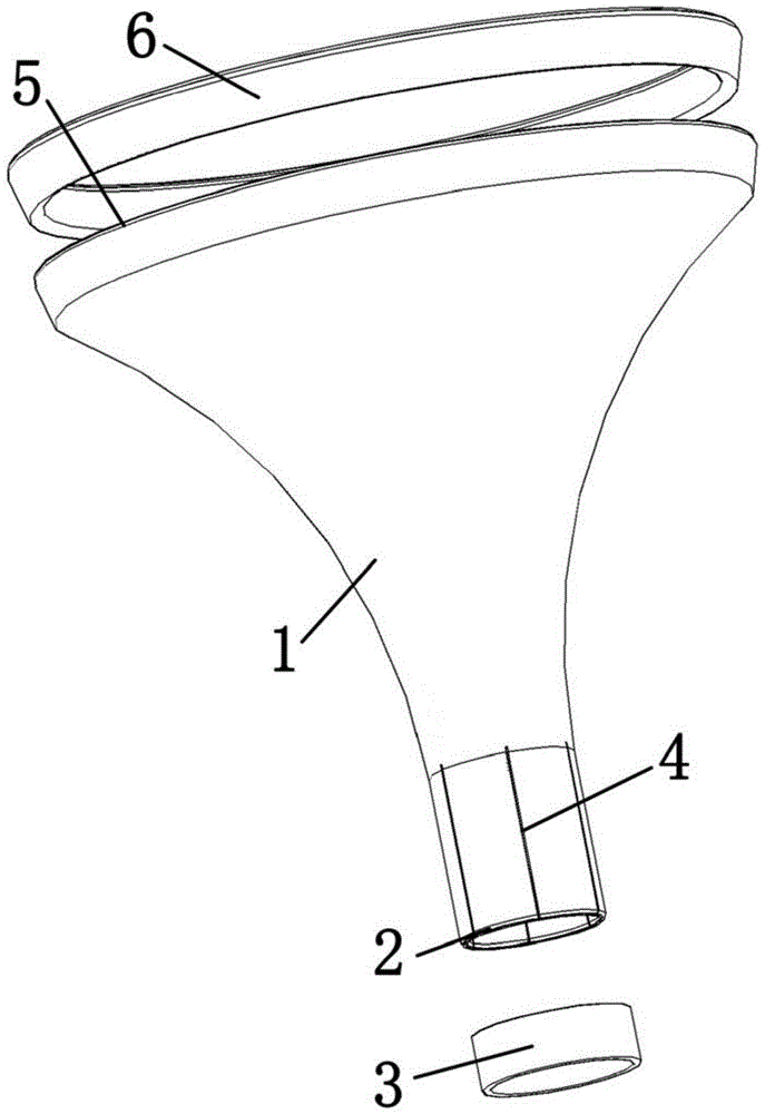
[0051] (2) Stamping and cutting

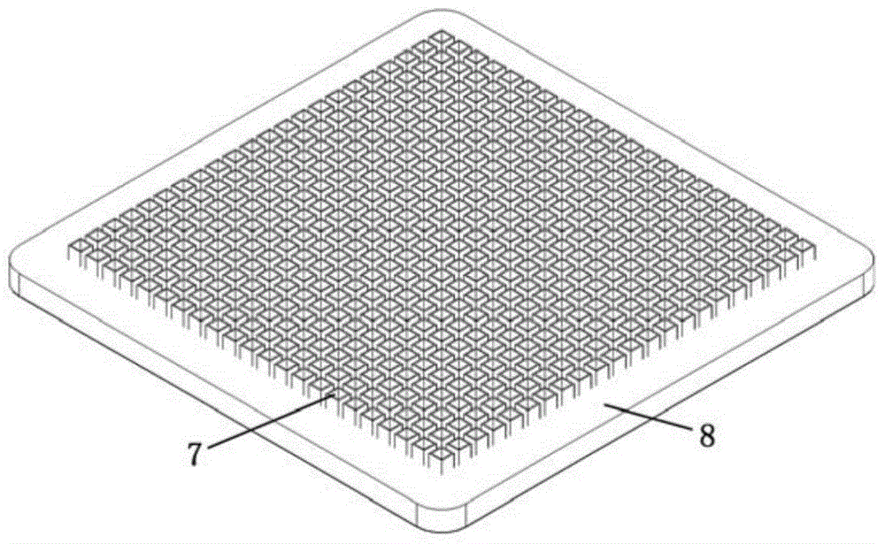
[0052] The amnion epithelium obtained in step (1) was quickly spread on the nitrocellulose filter membrane, and the amnion material obtained in step 1 was cut into a square amnion with a length of 3 mm and a width of 3 mm within 1 minute using a punching amnion cutting device Diaphragm;

[0053] (3) Compound thickening

[0054] Soak the amniotic membrane sheet prepared in step (2) in an aqueous solution of hyaluronic acid with a molecular weight of 1 million pharmaceutical grades at a concentration of 1% by mass for 1 minu...

Embodiment 2

[0059] A method for preparing a biofilm for glaucoma treatment, the steps of which are as follows:

[0060] (1)Quick collection of materials

[0061] The amniotic membrane rapid extraction device is used to quickly remove the amniotic membrane attached to the placenta around the umbilical cord within 3 minutes, and quickly refrigerate at 8°C, and transport it to the clean workshop for subsequent processing within 1 hour.

[0062] (2) Stamping and cutting

[0063] Spread the amnion epithelium obtained in step (1) upwards on the nitrocellulose filter quickly, and cut the amnion material obtained in step 1 into a rectangle with a length of 10 mm and a width of 3 mm within 3 minutes using a punching amnion cutting device Amniotic film.

[0064] (3) Compound thickening

[0065] Soak the amniotic membrane sheet prepared in step (2) in a pharmaceutical-grade high-viscosity sodium carboxymethylcellulose aqueous solution with a mass percent concentration of 5% for 3 minutes to thick...

Embodiment 3

[0070] A method for preparing a biofilm for glaucoma treatment, the steps of which are as follows:

[0071] (1)Quick collection of materials

[0072] The amniotic membrane rapid extraction device is used to quickly remove the amniotic membrane attached to the placenta around the umbilical cord within 2 minutes, and quickly refrigerate at 4°C, and transport it to the clean workshop for subsequent processing within 1 hour.

[0073] (2) Stamping and cutting

[0074] The amnion epithelium obtained in step (1) was quickly spread on the nitrocellulose filter membrane, and the amniotic membrane material obtained in step 1 was cut into circular amnion membrane pieces with a diameter of 6 mm within 2 minutes using a punching amnion membrane cutting device .

[0075](3) Compound thickening

[0076] Soak the amniotic membrane sheet prepared in step (2) in a 3% pharmaceutical-grade hyaluronic acid aqueous solution with a molecular weight of 1.5 million for 2 minutes to thicken the amni...


PUM

PropertyMeasurementUnit
thicknessaaaaaaaaaa
thicknessaaaaaaaaaa
thicknessaaaaaaaaaa

Abstract

The invention discloses a biofilm used for treating glaucoma and a preparation method thereof. The biofilm used for the treatment of glaucoma is thickened by soaking the amniotic membrane in an aqueous polymer polysaccharide solution with a concentration of 1-5%, and then sterilized by irradiation at -16-4°C, with a thickness of 0.2-0.5mm. The preparation method of the biofilm used for the treatment of glaucoma uses the thick amniotic membrane around the umbilical cord as a raw material, and undergoes the steps of rapid material collection, stamping and cutting, composite thickening, and irradiation sterilization. The beneficial technical effects of the present invention are: (1) various natural active ingredients of amniotic membrane can be effectively retained, including collagen, growth factors, anti-inflammatory factors, etc.; (2) biological activity equal to that of fresh amniotic membrane; (3) greatly improved (4) The thickness and degradation properties can fully meet the material requirements for subscleral flap filling in the treatment of glaucoma, and the biofilm can effectively play the role of preventing adhesion in the early stage after surgery and preventing scar formation in the later stage.

Description

[0001] 【Technical field】 [0002] The invention relates to the field of medical appliances, in particular to the technical field of tissue engineering Chinese medical biomaterials, in particular to a biofilm for glaucoma treatment and a preparation method thereof. [0003] 【Background technique】 [0004] Glaucoma is currently the second leading cause of blindness in the world, and the most commonly used surgical treatment is filtering surgery, but postoperative scarring of the filtering tract is often the main reason for surgical failure. There is no stable and good solution to this international problem. At present, there are glaucoma drainage devices with holes in sheet shape prepared by polymer materials such as polyethylene, polypropylene, polytetrafluoroethylene, or metal magnetic materials (such as the publication number is Chinese patent CN1337212A Chinese patent application, and the Chinese patent publication number is The Chinese patent application of CN103732182A, Ch...

Claims


Application Information

Patent Timeline
Patent Type & Authority Patents(China)
IPC IPC(8): A61L27/36A61L27/20A61L27/54
Inventor 张明昌刘艳丽刘博文王二浩何海娜叶伟初李翊翔林永亮谢丽君
Owner 广州瑞泰生物科技有限公司
Why Eureka
Try Eureka