A kind of preparation method of sofosbuvir crystal form 6
A technology of sofosbuvir crystal and sofosbuvir, which is applied in the field of medicine, can solve the problems of infeasibility, lengthy steps, difficult process control, etc., and achieves a simple and efficient crystallization process, good stability and good repeatability Effect
- Summary
- Abstract
- Description
- Claims
- Application Information
AI Technical Summary
Problems solved by technology
Method used
Image
Examples
preparation example Construction
[0042] In the preparation method of crystal form 6 of the present invention, sofosbuvir all uses the sofosbuvir product (such as J.Med.Chem.2010,53,7202-7218) prepared by the method reported in existing literature; Other All solvents and reagents were commercially available chemically pure or analytically pure products.
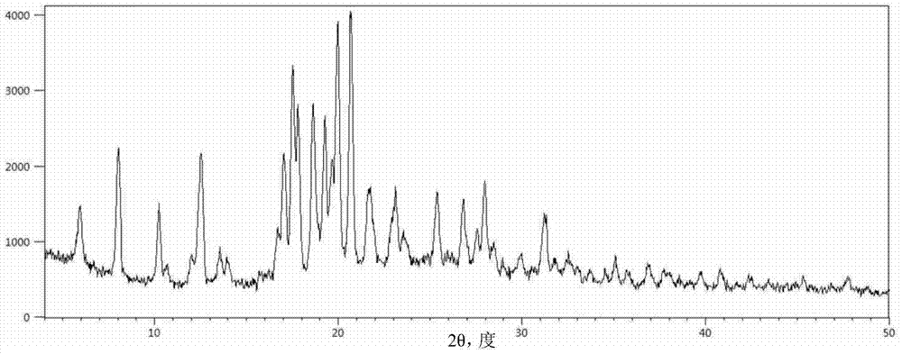
[0043] The X-ray powder diffractometer used in the embodiment of the present invention is an X'pert PRO X-ray powder diffractometer from PANalytical Company. Cu-Kα rays are used, the test power is 40kV×250mA, the scanning speed is 5° / min, and the scanning range is 4-80° (2θ) θ-2θ continuous scanning. In the X-ray powder diffraction diagram obtained in the embodiment of the present invention, the horizontal axis represents the 2θ position of the diffraction peak (unit: degree); the vertical axis represents the intensity of the diffraction peak.
[0044]The differential scanning calorimetry-thermogravimetric (DSC-TGA) analysis instrument used in the embodiment...
Embodiment 1
[0047] Mix 1.0 g of sofosbuvir with 10.0 ml of cyclopentyl methyl ether and 1.0 ml of anisole, heat the mixture to 75±3°C and stir at 20 rpm to completely dissolve the solids, then cool within 90 minutes to 10±3°C and stand at this temperature for 48 hours, the precipitated white powdery crystals were taken out by filtration, washed with 3 ml of isopropyl ether and dried to obtain 0.92 g of product.
[0048] Carry out powder X-ray diffraction analysis to the obtained crystal sample, its result is as follows figure 1 shown. and figure 1 Correspondingly, the powder X-ray diffraction data of Sofosbuvir Form 6 are shown in Table 1.
[0049] The pXRD characteristic peak position of table 1 embodiment 1 product
[0050]
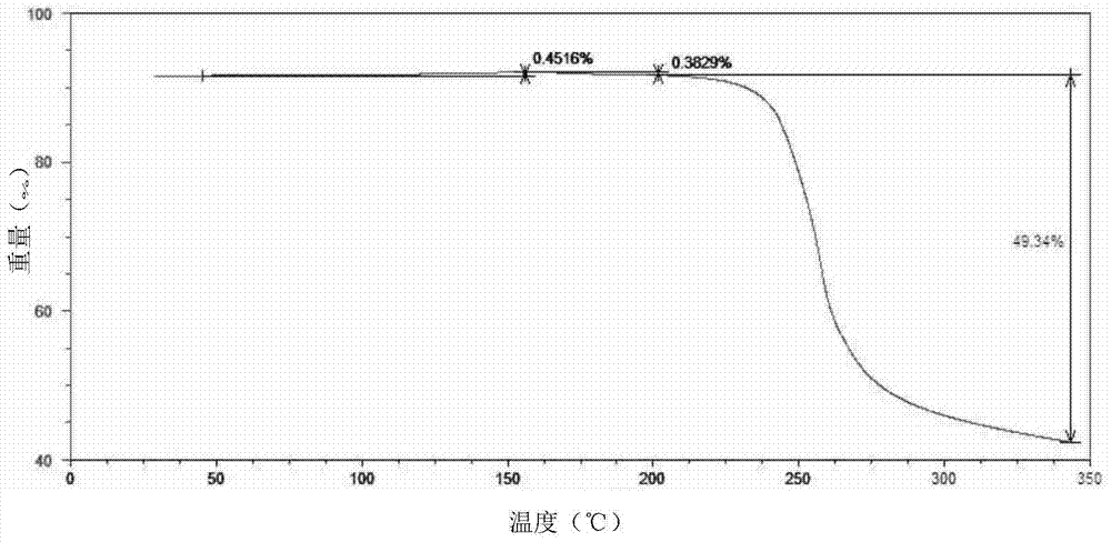
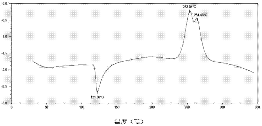
[0051] The powder sample was subjected to differential scanning calorimetry-thermogravimetric analysis, and the results were as follows: figure 2 shown. The shown pXRD, DSC-TGA and other test results are all the same as the characteristic spectrum of sofos...
Embodiment 2
[0053] Mix 1.0 g of sofosbuvir with 10.0 ml of cyclopentyl methyl ether and 2.0 ml of dibenzyl ether, heat the mixture to 75±3°C and stir at 20 rpm to completely dissolve the solid, then cool within 90 minutes to 5±3°C and stirred at this temperature for 48 hours, the precipitated white powdery crystals were taken out by filtration, washed with 3 ml of isopropyl ether and dried to obtain 0.93 g of the product with a yield of 93%. After the product was ground, pXRD, DSC-TGA and other tests were performed, all of which were identical to the characteristic spectrum of sofosbuvir crystal form 6 reported in the literature, confirming that the product was sofosbuvir crystal form 6.
PUM
 Abstract
Description
Claims
Application Information
 - R&D
- Intellectual Property
- Life Sciences
- Materials
- Tech Scout
- Unparalleled Data Quality
- Higher Quality Content
- 60% Fewer Hallucinations
Browse by: Latest US Patents, China's latest patents, Technical Efficacy Thesaurus, Application Domain, Technology Topic, Popular Technical Reports.
© 2025 PatSnap. All rights reserved.Legal|Privacy policy|Modern Slavery Act Transparency Statement|Sitemap|About US| Contact US: help@patsnap.com


