Probe and kit for detecting HER-2 (human epidermal growth factor receptor-2) gene amplification
A technology of HER-2 and gene amplification, which is applied in the determination/inspection of microorganisms, biochemical equipment and methods, DNA/RNA fragments, etc., can solve problems such as dependence, achieve high accuracy, reduce medical costs and expenses, The result is a stable effect
- Summary
- Abstract
- Description
- Claims
- Application Information
AI Technical Summary
Problems solved by technology
Method used
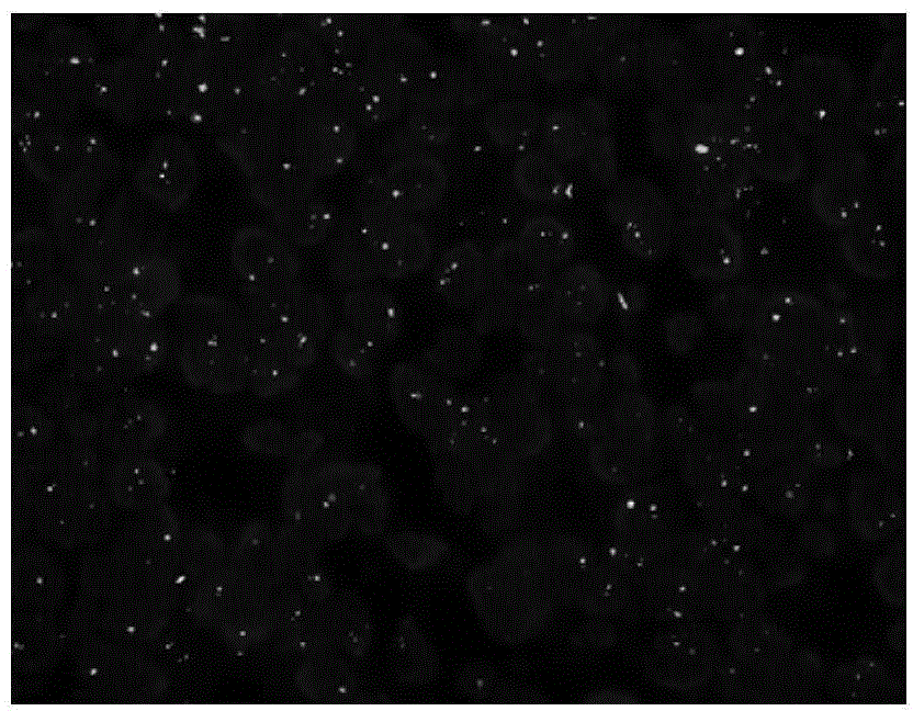
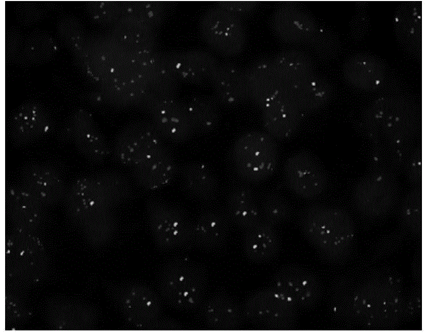
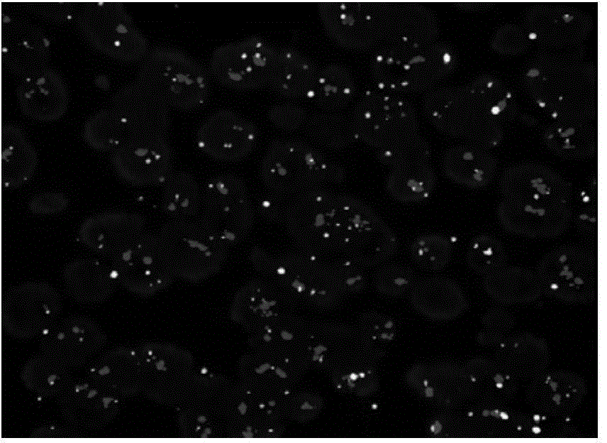
Image

Examples
Embodiment 1
[0024] Preparation of embodiment 1 HER-2 / CSP17 probe
[0025] Use the InvitrogenBioPrimeDNALabelingSystem (Invitrogen18094-011) kit to prepare HER-2 / CSP17 probes by random primer labeling, including the following process:
[0026] 1. Label the probe in a light-proof environment: take out the kit and related reagents from -20°C, thaw on ice, centrifuge briefly after thawing, and then put it back on ice. The probe labeling reference system is shown in Table 2.
[0027] Table 2 Probe Labeling Reference System
[0028] system
10 μL
20 μL
25 μL
N (40-80ng)
N (80-160ng)
N (100-200ng)
[0029] 2.5×Buffer
4μL
8μL
10 μL
dH 2 o
1-N
2-N
2.5-N
Lack of C / T dNTP
1.6μL
3.2 μL
4μL
Cy3-dCTP / FITC-dUTP
3.2 μL
6.4μL
8μL
Klenow Fragment (40U / μL)
0.2 μL
0.4μL
0.5μL
[0030] The HER-2 BACDNA used in the probe labeling is the BAC clone with th...
Embodiment 2
[0042] Embodiment 2 reagent preparation
[0043] (1) Preparation of pretreatment solution (pH7.0): weigh 22.6gNa 2 EDTA-2H 2 O and 6.34g Tris were dissolved in 800mL deionized water, adjusted to pH 7.0 after dissolving, and adjusted to 1L. Store in airtight at 2-8°C for 12 months. If the reagent becomes turbid or contaminated, please discard it immediately.
[0044] (2) Preparation of 20×SSC solution (pH7.0): weigh 88g of sodium citrate and 176g of NaCl in 800mL of deionized water, adjust the pH to 7.0 after dissolving, and dilute to 1L. Store in airtight at 2-8°C for 12 months. If the reagent becomes turbid or contaminated, please discard it immediately.
[0045] (3) Preparation of 2×SSC solution (pH7.0): Dilute 20×SSC solution 10 times with deionized water and adjust the pH to 7.0. Store in a sealed container at 2-8°C for 12 months. If the reagent is turbid or contaminated, please Discard immediately.
[0046] (4) Preparation of 20mg / mL proteinase K mother solution: Wei...
Embodiment 3
[0051] Application of a FISH kit for detection of HER-2 gene amplification in FFPE samples for detection of clinical samples
[0052] From February 2014 to August 2014, 80 paraffin-embedded tissue sections of clinical breast cancer were sent to our company for FISH detection of HER-2 gene amplification. Detection consists of the following steps:
[0053] 1. Pretreatment of FFPE samples, including the following steps:
[0054] (1) baked slices
[0055] (2) Xylene dewaxing
[0056] (3) Gradient ethanol hydration
[0057] (4) Pretreatment solution for 20 minutes
[0058] (5) Proteinase K digestion for 5-15min.
[0059] (6) Gradient ethanol dehydration step by step
[0060] 2. Under dark conditions, denature and hybridize the probe with the tissue to be tested on the glass slide, specifically including the following steps:
[0061] (1) Turn on the hybridization instrument, set or select the saved program: denaturation at 85°C for 5 minutes, hybridization at 37°C overnight (...
PUM
 Abstract
Description
Claims
Application Information
 - R&D
- Intellectual Property
- Life Sciences
- Materials
- Tech Scout
- Unparalleled Data Quality
- Higher Quality Content
- 60% Fewer Hallucinations
Browse by: Latest US Patents, China's latest patents, Technical Efficacy Thesaurus, Application Domain, Technology Topic, Popular Technical Reports.
© 2025 PatSnap. All rights reserved.Legal|Privacy policy|Modern Slavery Act Transparency Statement|Sitemap|About US| Contact US: help@patsnap.com