Three-dimensional cell assembly method based on dielectrophoresis adsorption principle

A three-dimensional assembly and dielectrophoresis technology, used in tissue regeneration, medical science, prosthesis, etc., to achieve the effect of high structural strength and precise control of cell distribution

Inactive Publication Date: 2016-06-22
BEIHANG UNIV
View PDF4 Cites 2 Cited by
  • Summary
  • Abstract
  • Description
  • Claims
  • Application Information

AI Technical Summary

Problems solved by technology

In addition to the characteristics of biocompatibility, degradability, and structural strength, the tissue engineering scaffold in this technolog

Method used


Image

  • Three-dimensional cell assembly method based on dielectrophoresis adsorption principle
  • Three-dimensional cell assembly method based on dielectrophoresis adsorption principle

Examples


Embodiment 1

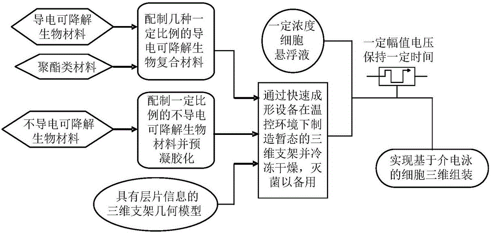
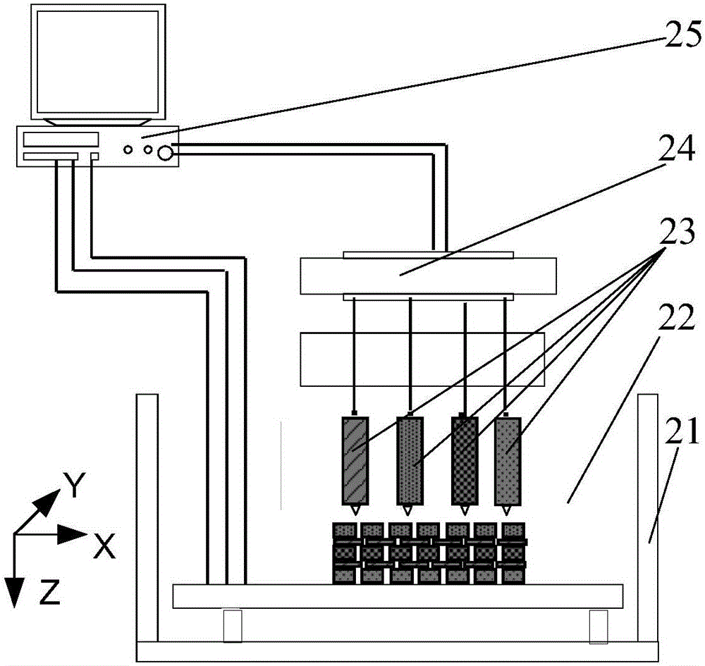
[0023] Example 1: Manufacturing a three-dimensional cartilaginous tissue structure by this method. Prepare gelatin into an aqueous solution with a concentration of 100g / L, which is called material A; dilute 50% glutaraldehyde with sterilized phosphate buffer solution 50 times to form a 1% glutaraldehyde solution, and filter it through a 0.22 μm filter membrane for later use. Call it material B; take the concentration as 1×10 6 / mL chondrocyte suspension 100mL spare, referred to as material C; the magnesium oxide-free particles wrapped by polypyrrole or poly(3,4-ethylenedioxythiophene)-polystyrenesulfonic acid and polylactic acid-glycolic acid copolymer (50:50) is prepared into a sol solution, and its weight and volume percentage concentration is 8%, which is called material D; material A and material D are respectively loaded into the material cavity and equipped with a nozzle; according to the pre-designed structure and definition planning Pathway, the above mixture is stacke...

Embodiment 2

[0024] Example 2: Manufacture a three-dimensional structure of bone-cartilage tissue by this method. Prepare gelatin into an aqueous solution with a concentration of 100g / L, which is called material A; dilute 50% glutaraldehyde with sterilized phosphate buffer solution 50 times to form a 1% glutaraldehyde solution, and filter it through a 0.22 μm filter membrane for later use. Call it material B; take the concentration as 5×10 6 / mL chondrocyte suspension 100mL spare, called material C1; the concentration is 1×10 6 / mL osteoblast suspension 100mL spare, called material C2; the magnesium oxide-free particles wrapped by polypyrrole or poly(3,4-ethylenedioxythiophene)-polystyrenesulfonic acid were copolymerized with polylactic acid-glycolic acid Material (50:50) is prepared into a sol solution, and its weight and volume percentage concentration is 8%, which is called material D; material A and material D are respectively loaded into the material cavity and equipped with a nozzle...

Embodiment 3

[0025] Example 3: The liver tissue-like three-dimensional structure of hepatic parenchymal cells-hepatic stromal cells-vascular endothelial cells was produced by this method. Prepare gelatin into an aqueous solution with a concentration of 100g / L, which is called material A; dilute 50% glutaraldehyde with sterilized phosphate buffer solution 50 times to form a 1% glutaraldehyde solution, and filter it through a 0.22 μm filter membrane for later use. Call it material B; take the concentration as 1×10 7 / mL liver parenchymal cell suspension 100mL spare, called material C1; the concentration is 2×10 6 / mL liver stromal cell suspension 100mL spare, called material C2; take the concentration of 5 × 10 6 / mL vascular endothelial cell suspension 100mL spare, called material C3; the magnesium oxide-free particles wrapped by polypyrrole or poly(3,4-ethylenedioxythiophene)-polystyrenesulfonic acid and polylactic acid-glycolic acid copolymerized Material (50:50) is prepared into a sol ...


PUM

PropertyMeasurementUnit
Layer thicknessaaaaaaaaaa

Abstract

The invention discloses a three-dimensional cell assembly method based on a dielectrophoresis adsorption principle and belongs to the technical field of artificial manufacture of tissue and organs. According to the method, firstly, a conductive and degradable biological material is prepared to serve as a scaffold main body conducting layer for cell assembly, a non-conductive material with good biocompatibility is taken as a scaffold insulating layer, an artificial tissue organ layer information model with the material area calibrated is established with a computer, thus the information model is used for directly driving multi-sprayer rapid forming equipment to manufacture a conductive three-dimensional tissue engineering scaffold, the conductive three-dimensional tissue engineering scaffold is freeze-dried, electrode pairs are mounted on the sterilized three-dimensional scaffold, the three-dimensional scaffold is immersed into a selected cell suspension, alternating current with a certain amplitude value is conducted for a period of time, and three-dimensional cell assembly is realized. On the basis of the conductive degradable biological material scaffold and the dielectrophoresis adsorption principle, three-dimensional assembly of one or more kinds of cells can be realized.

Description

technical field [0001] The invention discloses a three-dimensional cell assembly method based on the principle of dielectrophoretic adsorption, and belongs to the technical field of artificial manufacturing of tissues and organs. Background technique [0002] In the mid-1980s, LangerR, a chemical engineer at the Massachusetts Institute of Technology, and VacantiJP, an associate professor of surgery at Harvard Medical School, proposed the concept of tissue engineering (TissueEngineering). The essence of tissue engineering is that cells are combined in some way with matrix (Matrix) or carrier scaffold (Scaffold) to create new tissues. Tissue engineering has created a new way to overcome the many shortcomings of the previous clinical tissue and organ repair and reconstruction techniques, and has received widespread attention and rapid development all over the world. Tissue engineering research based on the combination of cells and carrier scaffolds. Specifically, the scaffolds...

Claims


Application Information

Patent Timeline
IPC IPC(8): A61L27/38A61L27/22A61L27/18A61L27/20A61L27/58A61L27/02
CPCA61L27/3808A61L27/025A61L27/18A61L27/20A61L27/222A61L27/3804A61L27/3817A61L27/3821A61L27/3826A61L27/3839A61L27/3847A61L27/3852A61L27/3886A61L27/58A61L2430/02A61L2430/06A61L2430/20A61L2430/28C08L89/06C08L79/04C08L65/00C08L67/04C08L5/08C08L5/04
Inventor 刘海霞梁振江戴向国刘凯铭张忠钢
Owner BEIHANG UNIV
Why Eureka
Try Eureka