Method for extracting genomic DNA (deoxyribonucleic acid) from whole blood on basis of improved immunomagnetic bead method

A genome and magnetic bead method, applied in the biological field, can solve the problems of large amount of reagents, long test time, low qualification rate, etc., and achieve the effect of small amount of reagents, reduced test cost and short time.

Inactive Publication Date: 2016-07-13
武汉血液中心
View PDF4 Cites 3 Cited by
  • Summary
  • Abstract
  • Description
  • Claims
  • Application Information

AI Technical Summary

Problems solved by technology

[0002] There are many methods for extracting DNA. The traditional phenol chlorine method, salting-out method, and column-passing method can only be extracted manually and cannot be automated. The magnetic bead method uses the specific adsorption of cellulose wrapped with magnetic beads to DNA. Separation and acquisition of DNA under the action of dehydration is easy to automate. The DNA magnetic bead extraction method has many advantages, but the concentration and purity of DNA extracted by this method are easily affected by the residual amount of magnetic beads, lysis time, whole blood volume and enzyme volume. These factors have an impact through interaction, resulting in unstable DNA concentration and low purity, which in turn affect HLA typing results. High, long test time

Method used


Image

  • Method for extracting genomic DNA (deoxyribonucleic acid) from whole blood on basis of improved immunomagnetic bead method
  • Method for extracting genomic DNA (deoxyribonucleic acid) from whole blood on basis of improved immunomagnetic bead method
  • Method for extracting genomic DNA (deoxyribonucleic acid) from whole blood on basis of improved immunomagnetic bead method

Examples


Embodiment 1

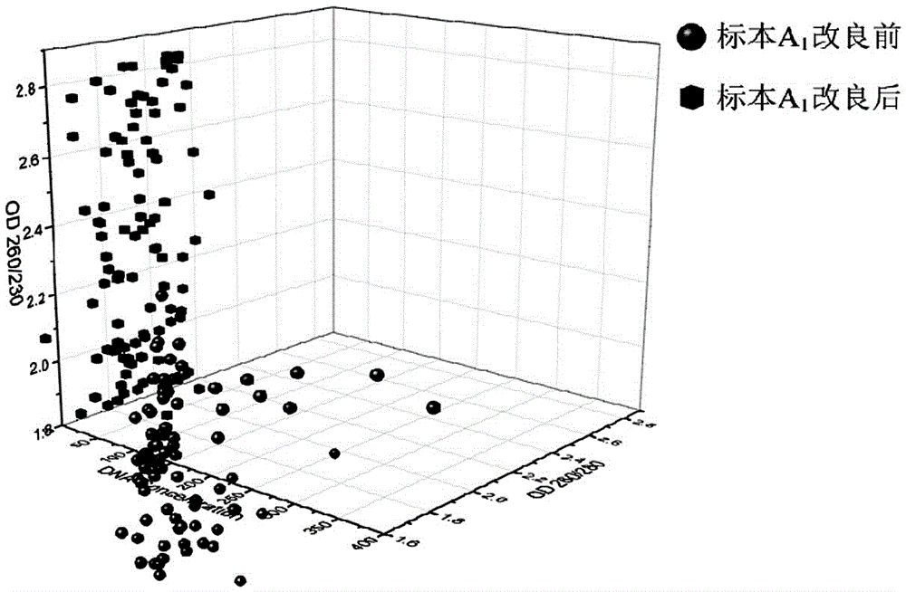
[0028] Whole blood sample A 1 Blood samples from stem cell donors in Hubei Branch of China Bone Marrow Bank.

[0029] Utilize the method of the present invention to above-mentioned whole blood sample specimen A 1 DNA detection, the specific steps are as follows:

[0030] (1) Red blood cell lysis

[0031] Add 500 μl of red blood cell lysate into a 2ml centrifuge tube, add 120 μl of completely thawed whole blood sample into the above centrifuge tube, oscillate fully, and mix until the cell fragments are evenly suspended and no clumps are seen. Dilute the lysate × 10,000 Rmp was centrifuged for 1 min, and the supernatant was removed.

[0032] (2) Leukocyte Lysis and DNA Adsorption

[0033] Dilute the proteinase K stock solution to a ratio of two times, draw 20 μl proteinase K and 140 μl LysisBuffer into the above centrifuge tube, and repeatedly pipette and mix 3 times, then incubate at 65°C, shake for 2 minutes and rest for 8 minutes, and alternately shake and stand for 3 cyc...

Embodiment 2

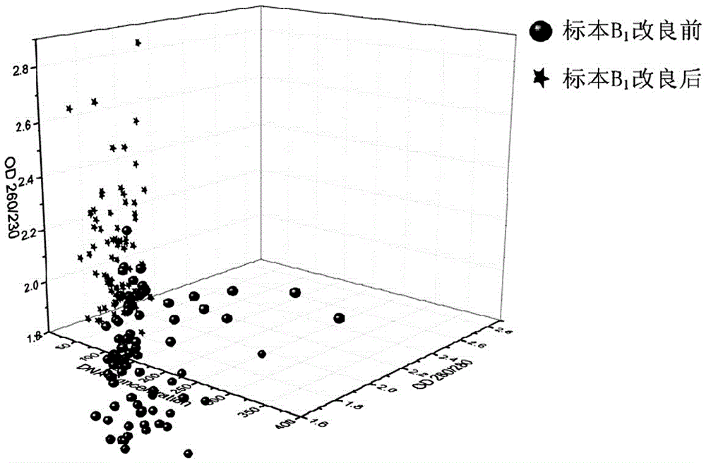
[0048] Whole blood sample B 1 Blood samples from stem cell donors in Hubei Branch of China Bone Marrow Bank.

[0049]Utilize the method of the present invention to above-mentioned whole blood sample specimen B 1 DNA detection, the specific steps are as follows:

[0050] (1) Red blood cell lysis

[0051] Add 480 μl of red blood cell lysate into a 2ml centrifuge tube, add 130 μl of completely thawed whole blood sample into the above centrifuge tube, oscillate fully, and mix until the cell fragments are evenly suspended and no clumps are seen. Dilute the lysate × 10,000 Rmp was centrifuged for 2min, and the supernatant was removed.

[0052] (2) Leukocyte Lysis and DNA Adsorption

[0053] Dilute the protease K stock solution in multiple ratios, pipette 20 μl proteinase K and 150 μl LysisBuffer into the above centrifuge tube, and repeatedly pipette and mix 3 times, then incubate at 65°C, shake for 2.5 minutes and rest for 6 minutes, and alternately shake and rest for 3 cycles; ...

Embodiment 3

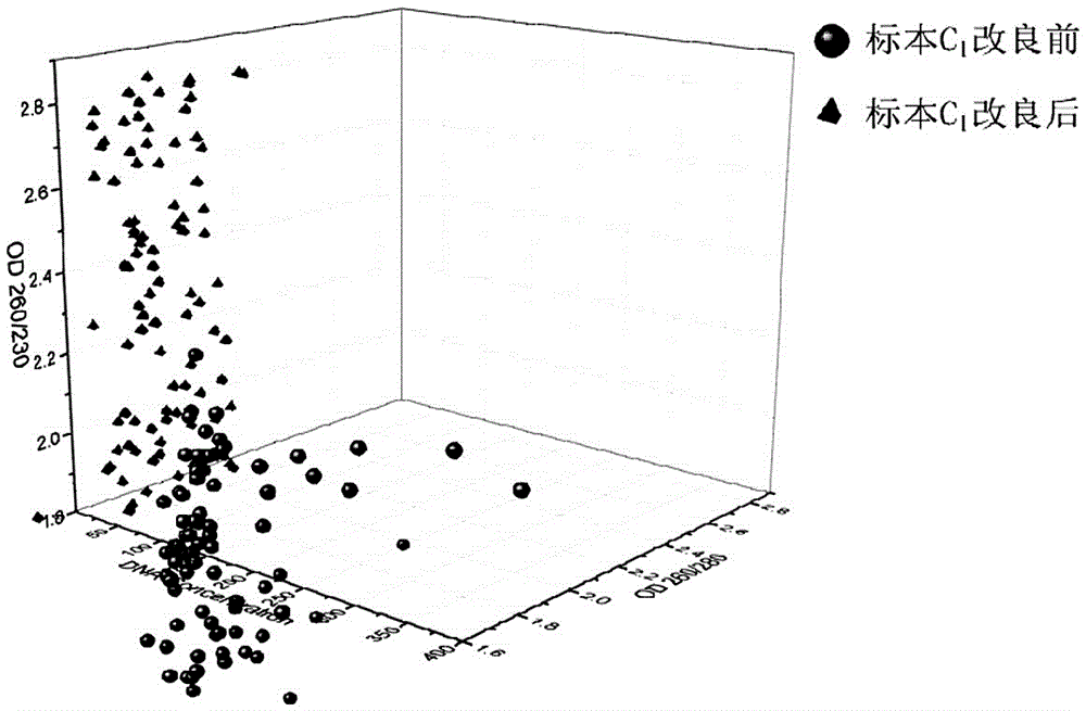
[0068] Whole blood sample C 1 Blood samples from stem cell donors in Hubei Branch of China Bone Marrow Bank.

[0069] Utilize the method of the present invention to above-mentioned whole blood sample C 1 DNA detection, the specific steps are as follows:

[0070] (1) Red blood cell lysis

[0071] Add 450 μl of red blood cell lysate into a 2ml centrifuge tube, add 150 μl of completely thawed whole blood sample into the above centrifuge tube, oscillate fully, and mix until the cell fragments are evenly suspended and no clumps are seen. Dilute the lysate × 10,000 Rmp was centrifuged for 3min, and the supernatant was removed.

[0072] (2) Leukocyte Lysis and DNA Adsorption

[0073] Dilute the proteinase K stock solution to a ratio of two times, draw 20 μl proteinase K and 120 μl LysisBuffer into the above centrifuge tube, and repeatedly pipette and mix 3 times, then incubate at 65°C, shake for 3 minutes and rest for 5 minutes, and alternately shake and stand for 3 cycles; add 3...


PUM


Abstract

The invention relates to a method for extracting genomic DNA (deoxyribonucleic acid) from whole blood on basis of an improved immunomagnetic bead method.The method includes steps: erythrocyte splitting, leukocyte splitting, DNA adsorption, DNA purification and DNA elution.DNA concentration and purity are measured by a DNA quantometer, sequencing of PCR (polymerase chain reaction) amplification products is realized by a sequencer, and HLA (human leukocyte antigen) classification result analysis is performed through relevant analysis software.The method has the advantages of high DNA concentration uniformity, high purity, substantial increase of HLA classification result success rate, and high acceptability of a signal-to-noise ratio, thereby being widely applicable to extraction of the genomic DNA from whole blood samples.

Description

technical field [0001] The invention belongs to the field of biotechnology, and in particular relates to a method for rapidly extracting genomic DNA in whole blood based on an improved magnetic bead method. Background technique [0002] There are many methods for extracting DNA. The traditional phenol chlorine method, salting-out method, and column-passing method can only be extracted manually and cannot be automated. The magnetic bead method uses the specific adsorption of cellulose wrapped with magnetic beads to DNA. Separation and acquisition of DNA under the action of dehydration is easy to automate. The DNA magnetic bead extraction method has many advantages, but the concentration and purity of DNA extracted by this method are easily affected by the residual amount of magnetic beads, lysis time, whole blood volume and enzyme volume. These factors have an impact through interaction, resulting in unstable DNA concentration and low purity, which in turn affect HLA typing r...

Claims


Application Information

Patent Timeline
Patent Type & Authority Applications(China)
IPC IPC(8): C12N15/10
CPCC12N15/1013
Inventor 李王霞沈钢李刚姚立马严仇丰武江梦天刘光箭邹洁朱远雁
Owner 武汉血液中心
Why Eureka
Try Eureka