Mitochondria injection and application thereof

A technology of mitochondria and injection, applied in the field of biomedicine to achieve the effect of improving cell activity and increasing ATP production

Inactive Publication Date: 2016-07-20
SOUTHWEST UNIVERSITY
View PDF0 Cites 4 Cited by
  • Summary
  • Abstract
  • Description
  • Claims
  • Application Information

AI Technical Summary

Problems solved by technology

[0004] However, there is no preparation of mitochondria into pharm

Method used


Image

  • Mitochondria injection and application thereof
  • Mitochondria injection and application thereof
  • Mitochondria injection and application thereof

Examples


Embodiment 1

[0028] Example 1. Preparation of mitochondrial intravenous injection for the treatment of drug-induced liver injury

[0029] Extraction of liver mitochondria: liver tissue was taken, and the animal was fasted for 12 hours before sampling, then quickly decapitated on ice, and 0.15 g of tissue was cut into 0.5 cm 2 Fragments, washed with washing solution (1mM Tris-HCl, pH7.0, 0.13MNaCl, 5mMKCl and 7.5mMMgCl 2 ) after washing, centrifuge at 800g for 10min; separate liquid (4mM Tris-HCl, pH7.4, 2.5mMNaCl and 0.5mMMgCl 2 ) resuspended, and allowed to stand for 1 to 2 minutes to collect the cells; crush the collected cells with a homogenizer to obtain a homogenate; transfer the homogenate to a centrifuge tube, and centrifuge at 4°C and 800×g for 5 minutes; collect Transfer the supernatant to a new centrifuge tube, centrifuge again at 4°C, 800×g for 5 minutes, and discard the precipitate; then transfer the supernatant to a new centrifuge tube, centrifuge at 4°C, 10,000×g for 10 minu...

Embodiment 2

[0036] Embodiment 2, preparation for treating Parkinson's disease mitochondrial injection

[0037] Extraction of brain mitochondria: take normal animal brain, homogenize with a glass homogenizer, transfer the homogenate to a centrifuge tube, centrifuge at 800×g for 5 min, collect the supernatant into a new centrifuge tube; transfer the supernatant to Centrifuge a new centrifuge tube at 10000×g for 10 min; then add 0.2 mL of mitochondrial extraction and purification solution (225 mM mannitol, 75 mM sucrose, 10 mM KCl, 10 mM Tris and pH7.0, 5 mM KH 2 PO 4 ) to resuspend the mitochondrial pellet, and centrifuge at 12000×g for 10 min; discard the supernatant, and the pellet is the mitochondria.

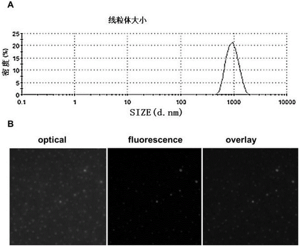
[0038] Mitochondria modification: disperse the extracted mitochondria with normal saline, slowly add the chitosan solution with a mass fraction of 2% to the mitochondrial solution at a mass ratio of 1:5, shake well, and store at room temperature (18-25°C) After standing for 5 minutes, a...

Embodiment 3

[0041] Embodiment 3, the application of mitochondrial injection

[0042] In order to verify that the mitochondrial injection prepared by the present invention can be used for intravenous injection, and can treat mitochondria-related diseases, the mitochondrial injection was subjected to the following experiments:

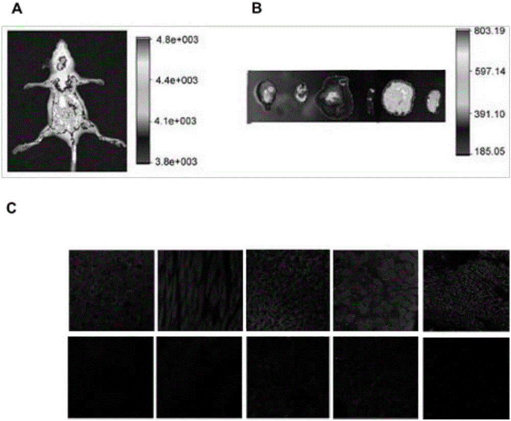
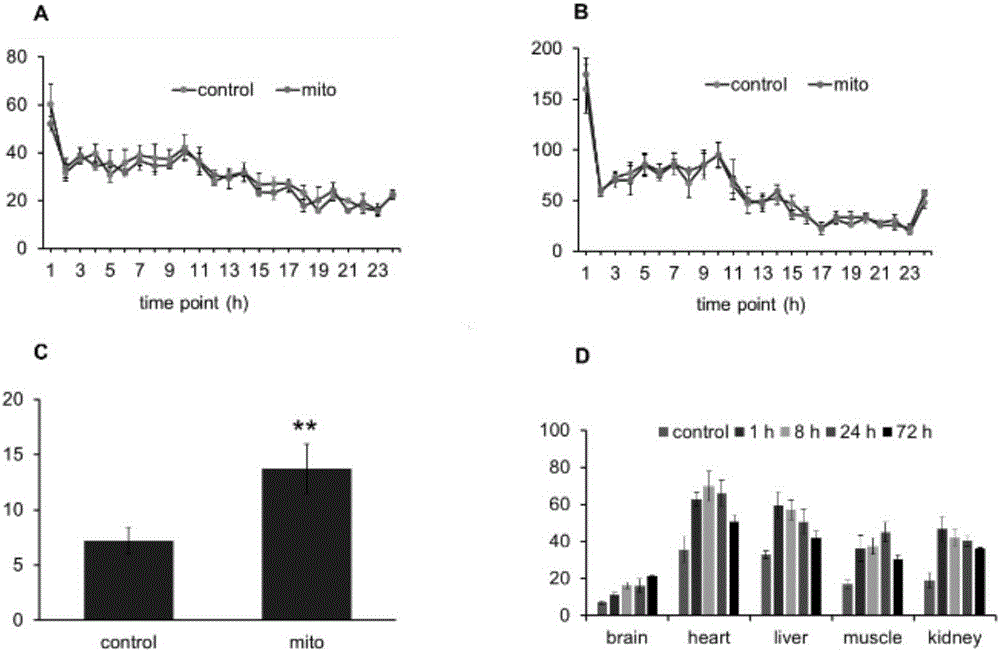
[0043] 1. The distribution of mitochondria in the body after intravenous injection

[0044] The mitochondrial injection prepared with normal saline (mitochondria were prepared for fluorescent staining) was injected into nude mice (0.5 mg / kg). After 2 hours of injection, the nude mice were anesthetized with 4% chloral hydrate by mass fraction and placed in Fluorescent imaging was performed in a small animal in vivo imager (Carestream Co.). Subsequently, the mice were perfused with ice-cold saline, and brain, liver, kidney, lung, skeletal muscle and other tissues were separated for tissue imaging. The results showed that there was a wide distribution of fluorescence ...


PUM

PropertyMeasurementUnit
Particle sizeaaaaaaaaaa
The average particle sizeaaaaaaaaaa

Abstract

The invention discloses a mitochondria injection and application thereof. The mitochondria injection is an injectable solution, an emulsion or a suspension prepared by adding solvents or other auxiliaries into mitochondria which is extracted from tissues or cells of animals, plants and microorganisms through performing germfree operation and the gradient centrifugation technology and serves as a raw material. The prepared mitochondria injection can be applied to local drug delivery and intracardial injection. After the injection is applied, the ATP content in the tissues can be increased, and the activity of the cells can be enhanced; the mitochondria injection is used for treating Parkinson's disease relevant to mitochondria damage and diseases such as liver injury induced by medicines, so that an effective and innovative treating method is provided for mitochondria-associated diseases.

Description

technical field [0001] The invention belongs to the field of biomedicine, and in particular relates to a mitochondrial intravenous injection, and also relates to the application of the mitochondrial injection preparation. Background technique [0002] Mitochondria, a multifunctional organelle in cells, are not only cellular energy (ATP) generation centers but also signaling hubs regulating reactive oxygen species, cell survival, and death. Mitochondrial diseases are a series of diseases related to mitochondrial dysfunction, including neurological and muscle degenerative diseases (mitochondrial muscle encephalopathy, mitochondrial myopathy, Parkinson's disease, Huntington's disease, etc.), aging, diabetes, obesity, cardiovascular disease, Drug-induced liver injury, etc. Mitochondrial dysfunction is a common cause of mitochondria-related diseases. At present, there is no fundamental treatment for this disease caused by mitochondrial dysregulation. [0003] Two important fac...

Claims


Application Information

Patent Timeline
IPC IPC(8): A61K35/30A61K35/407A61K35/12A61K35/56A61K35/66A61K36/00A61K9/08A61K9/10A61K9/107A61K47/36A61K47/34A61P25/28A61P21/00A61P9/00A61P13/12A61P3/10A61P35/00A61P39/06A61P3/04A61P1/16A61P25/16A61P25/14
CPCA61K9/0019A61K9/08A61K9/10A61K9/107A61K35/12A61K35/30A61K35/407A61K35/56A61K35/66A61K36/00A61K47/34A61K47/36
Inventor 付爱玲付琛史现勋赵明
Owner SOUTHWEST UNIVERSITY
Why Eureka
Try Eureka