Detection method for content of adenosine in biological sample

A technology for biological samples and adenosine content, applied in the field of high-sensitivity adenosine detection, can solve the problems of time-consuming operation and expensive instruments, and achieve the effect of wide linear range and high-sensitivity detection

Inactive Publication Date: 2016-08-17
NANJING MEDICAL UNIV
View PDF2 Cites 14 Cited by
  • Summary
  • Abstract
  • Description
  • Claims
  • Application Information

AI Technical Summary

Problems solved by technology

Although the current detection methods of adenosine can meet the detection requirements, there are many shortcomings, such as high performance liquid chromatography, ultraviolet detection, flow detection and gas/liquid chromatography,

Method used


Image

  • Detection method for content of adenosine in biological sample
  • Detection method for content of adenosine in biological sample
  • Detection method for content of adenosine in biological sample

Examples


Embodiment 1

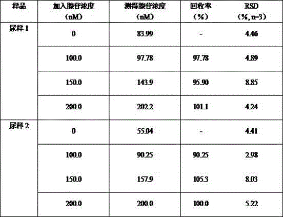
[0027] In this embodiment, the detection is the adenosine content in the urine of lung cancer patients, and the steps are as follows:

[0028] (1) Preparation of ferroferric oxide magnetic nanoparticles with carboxyl groups on the surface (P. Sun, H. Y. Zhang, C. Liu, J. Fang, Langmuir 2010, 26(2), 1278–1284.): Co-precipitation method Preparation of magnetic Fe 3 o 4 Nanoparticles: Add 80 mL deoxygenated ultrapure water into a 250 mL three-neck flask, stir magnetically, keep the stirring speed at 800 rpm, add 1.72 g of FeCl under nitrogen protection 2 · 4H 2 O and 4.72 g FeCl 3 · 6H 2 O, when the temperature rises to 80°C, add 10 mL of concentrated ammonia water and 2 mL of PEG dropwise, react for 1 h, wash with deionized water until neutral, and disperse in 100 mL of ethanol to obtain Fe 3 o 4 magnetic nanoparticles. Dissolve 10.5 g of citric acid monohydrate supernatant in 100 mL of water, add finely ground Fe 3 o 40.5 g, ultrasonically dispersed for 20 min, mecha...


PUM

PropertyMeasurementUnit
Particle sizeaaaaaaaaaa

Abstract

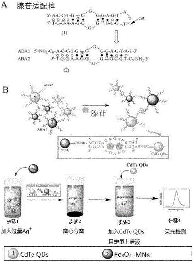
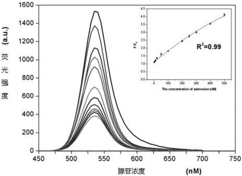
The invention relates to a detection method for the content of adenosine in a biological sample. An adenosine probe is formed by mixing ferriferrous oxide magnetic nano-particles with surfaces modified by an adenosine aptamer segment 1 and cadmium telluride quantum dots with surfaces modified with an adenosine aptamer segment 2; the adenosine probe and the adenosine are paired by an alkali group to form an adenosine sandwiched structure, and the adenosine sandwiched structure is separated from the biological sample; the content of the adenosine is quantified by applying ion exchange effect of silver ions and cadmium ions in the cadmium telluride quantum dots in the adenosine sandwiched structure; the method has the characteristics of very high sensitivity, accuracy and precision.

Description

technical field [0001] The invention belongs to the field of biological detection, and in particular relates to a highly sensitive adenosine detection method based on nucleic acid aptamer recognition technology and ion exchange signal amplification mechanism. Background technique [0002] Adenosine (AD) is adenosine nucleoside, which is a metabolite of RNA in the body and belongs to small biological molecules. In the central nervous system, it plays an important role in the regulation of neurotransmission and in resisting ischemic and disease-induced nerve injury. At the same time, adenosine is the substrate of adenosine deaminase, and its content change will also indirectly reflect the metabolic level of adenosine deaminase in vivo, and the detection of adenosine deaminase activity is an important reference for the diagnosis of many clinical diseases Therefore, the development of new detection methods for adenosine is of great significance for medical clinical diagnosis an...

Claims


Application Information

Patent Timeline
IPC IPC(8): G01N21/64G01N33/68
CPCG01N21/6428G01N33/6893
Inventor 胡琴宋越月
Owner NANJING MEDICAL UNIV
Why Eureka
Try Eureka