GRPR targeted molecular probe and preparation method thereof

A molecular probe and targeting technology, applied in the field of GRPR targeting molecular probe and its preparation, can solve the problems of low tumor/background contrast, low tumor uptake rate, poor pharmacokinetics, etc. To achieve good pharmacokinetic properties and biological distribution characteristics, accurate test results, and ensure the effect of accuracy

Inactive Publication Date: 2016-10-26
宁波益格爱生物科技有限公司
View PDF3 Cites 5 Cited by
  • Summary
  • Abstract
  • Description
  • Claims
  • Application Information

AI Technical Summary

Problems solved by technology

However, for clinical applications, such molecular probes generally have disadvantages such as low tumor uptake rate, low tumor / background contrast, mainly metabolized by the kidneys, and poor pharmacokinetics.

Method used


Image

  • GRPR targeted molecular probe and preparation method thereof
  • GRPR targeted molecular probe and preparation method thereof
  • GRPR targeted molecular probe and preparation method thereof

Examples


Embodiment 1

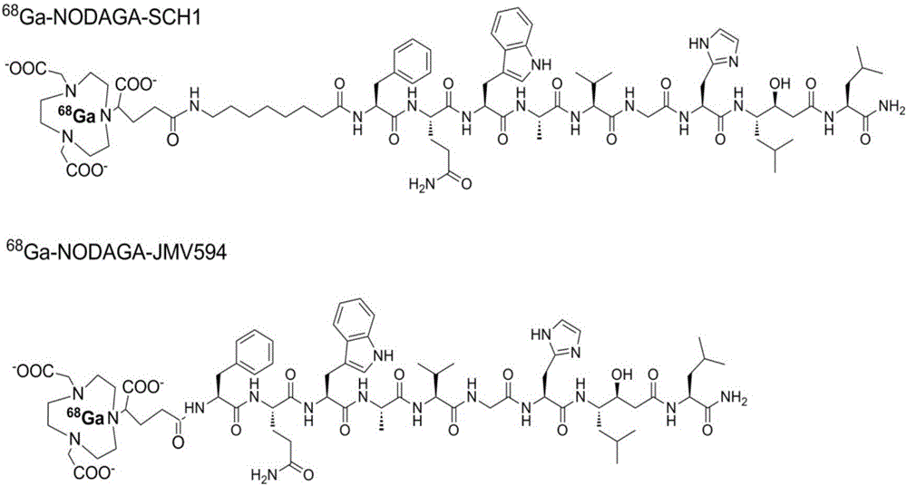
[0049] Embodiment 1: synthetic compound 68 Ga-NODAGA-JMV594

[0050]

[0051] The polypeptide JMV594 (1.13 mg, 0.01 mmol) and NODAGA-NHS ester (0.95 mg, 0.02 mmol, 2.0 equivalents) were weighed in a 2 mL test tube and dissolved in DMF solvent, and 2 equivalents of DIPEA were added. After 4 hours of reaction at room temperature. The reaction solution was directly purified by preparative HPLC and then vacuum freeze-dried to obtain a white solid powder NODAGA-JMV594, 0.80 mg, yield 55%. MS calculated value: C 70 h 103 N 17 o 18 + ([M+H] + ): 1470.8, measured value: ESI-MS: m / z 1470.8.

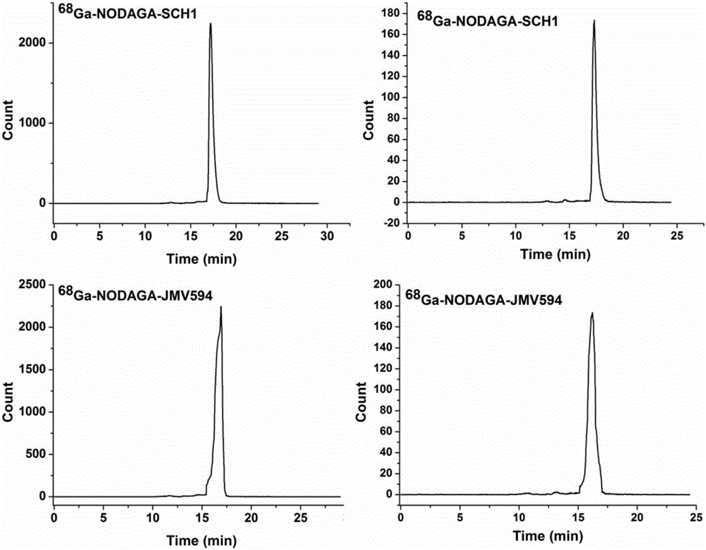
[0052] Dissolve 2nmol of NODAGA-JMV594 compound in 1mL NaOAc buffer (pH=4.5) and add 2mCi nuclides at 37°C 68 Ga labeling reaction 15min. labeled compound 68 After Ga-NODAGA-JMV594 was purified by HPLC, the organic solvent acetonitrile was blown dry with nitrogen gas, and diluted with PBS solution, it was directly used in cell uptake experiments and in vivo imaging experiments.

Embodiment 2

[0053] Embodiment 2: synthesis 68 Ga-NODAGA-SCH1

[0054]

[0055] Peptide SCH1 (1.25 mg, 0.01 mmol) and NODAGA-NHS ester (0.95 mg, 0.02 mmol, 2.0 equivalents) were weighed in a 2 mL test tube and dissolved in DMF solvent, and 2 equivalents of DIPEA were added. After 2 hours of reaction at room temperature. The reaction solution was directly purified by preparative HPLC and then vacuum freeze-dried to obtain a white solid powder NODAGA-SCH1, 1.30 mg, with a yield of 80%. MS calculated value: C 78 h 119 N 18 o 19 + ([M+H] + ): 1611.9, measured value: MALDI-TOF-MS: 1611.9.

[0056] Dissolve 2nmol of NODAGA-SCH1 compound in 1mL NaOAc buffer (pH=4.5) and add 2mCi nuclides at 37°C 68 Ga labeling reaction 15min. labeled compound 68 After Ga-NODAGA-SCH1 was purified by HPLC, the organic solvent acetonitrile was blown dry with nitrogen gas, and diluted with PBS solution, it was directly used in cell uptake experiments and in vivo imaging experiments.

Embodiment 3

[0057] Embodiment 3: synthetic compound 68 Ga-NODAGA-ZHS1

[0058]

[0059] Peptide ZH1 (1.28 mg, 0.01 mmol) and compound C1a (0.56 mg, 0.02 mmol, 2.0 equivalents) were weighed in a 2 mL test tube and dissolved in DMF solvent. After 2 hours of reaction at room temperature. The reaction solution was directly purified by preparative HPLC and then vacuum freeze-dried to obtain a white solid powder ZHS1, 1.13 mg, with a yield of 75%. MS calculated value: C 74 h 111 N 18 o 16 + ([M+H] + ): 1506.8, measured value: ESI-MS: 1506.9.

[0060] Weigh the polypeptide ZHS1 (1.56mg, 0.01mmol) and NODAGA-NHS ester (0.95mg, 0.02mmol, 2.0eq) in a 2mL test tube and dissolve them in DMF solvent, and add 2equivalents of DIPEA. After 4 hours of reaction at room temperature. The reaction solution was directly purified by preparative HPLC and then vacuum freeze-dried to obtain a white solid powder NODAGA-ZHS1, 1.50 mg, with a yield of 80%. MS calculated value: C 89 h 134 N 21 o 23 ...


PUM


Abstract

The invention belongs to the field of diagnostic imaging and relates to a GRPR targeted molecular probe and a preparation method thereof. The general formula of the GRPR targeted molecular probe is M-X-JMV594, wherein M is a signal component, X is a connector, and JMV594 is the targeted affinity component of GRPR; the JMV594 is a polypeptide sequence D-Phe-Gln-Trp-Ala-Val-Gly-His-Sat-Leu-NH2, and the signal component M is the combination of radioactive isotope with one or more of metal chelating agent, fluorescent dye, quantum dots, paramagnetic materials, super-paramagnetic materials, magnetic nano particles, ultrasonic microbubbles and photo-acoustic nano particles. The GRPR targeted molecular probe can be used as an image developing agent or radiotherapy medicine, has a good pharmacokinetics feature and a good biology distribution feature, and is accurate in detection result and promising in clinical application prospect.

Description

technical field [0001] The invention belongs to the field of diagnostic imaging agents, in particular to a GRPR targeting molecular probe and a preparation method thereof. Background technique [0002] Commonly used molecular imaging techniques include magnetic resonance imaging (MRI), computed tomography (CT), positron emission tomography (PET), and single photon emission computed tomography (single photon emission tomography). emission computed tomography, SPECT), optical imaging (including multiphoton imaging, near-infrared first region imaging, near infrared second region imaging and confocal imaging, etc.) and ultrasound imaging. Each imaging technique has its own advantages and disadvantages in terms of sensitivity, principle, resolution and acquisition time. [0003] Table 1 Comparison of various imaging modes [0004] [0005] The core of molecular imaging is to obtain high-quality images of specific target molecules in real time, so it needs to design various m...

Claims


Application Information

Patent Timeline
Patent Type & Authority Applications(China)
IPC IPC(8): A61K49/00A61K51/08A61K51/04A61K49/22A61K49/14C07K7/02
CPCA61K49/0002A61K49/0056A61K49/14A61K49/223A61K51/0474A61K51/0482A61K51/08C07K7/02
Inventor 程震
Owner 宁波益格爱生物科技有限公司
Why Eureka
Try Eureka